This is default featured slide 1 title

Go to Blogger edit html and find these sentences.Now replace these sentences with your own descriptions.

This is default featured slide 2 title

Go to Blogger edit html and find these sentences.Now replace these sentences with your own descriptions.

This is default featured slide 3 title

Go to Blogger edit html and find these sentences.Now replace these sentences with your own descriptions.

This is default featured slide 4 title

Go to Blogger edit html and find these sentences.Now replace these sentences with your own descriptions.

This is default featured slide 5 title

Go to Blogger edit html and find these sentences.Now replace these sentences with your own descriptions.

Sunday, February 26, 2006

Alopecia with peculiar regrowth pattern

The patient is a 35 year old healthy man who presented with alopecia of the scalp of 1 year duration. Many topical therapies were tried with no response. On examination, there was classical alopecia areata. I gave him IM triamcinolone 40mg injection every 2 weeks with topical Dermovate oinment for 2 months. He came back with hair growth in a wavy pattern in the form of concentric circles (see Figure).. I do not have an explanation. I would appreciate any helpful comments.


Presented by Professor Khalifa Sharquie
University of Baghdad, Iraq

Saturday, February 18, 2006

From the Nail File

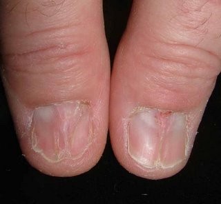
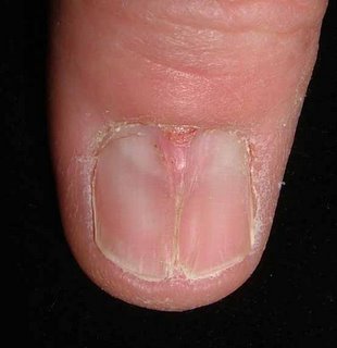
55 year old man with 6 month history of nail distrophy. Only thumbs. He works with his hands cutting film. Trauma likely.

The patient wrote:
"I noticed left thumbnail change in appearance in late August 2005. I scraped the nail down and got fluid near base. The right thumbnail became abnormal after trauma in October of 2005 showing a crack straight up the middle of the nail."


Moderator: I suspect lichen planus. Median nail dystrophy less likely.

Comments??

Saturday, February 11, 2006

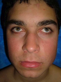
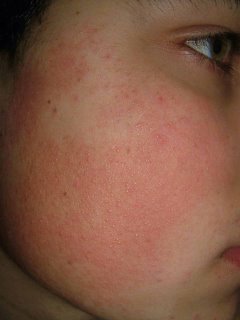
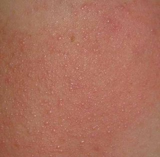
Erythromelanosis Follicularis Facei

The patient is a 15 year old boy with a many year history of ruddy cheeks and a fine follicular eruption in the affected areas. He also has typical keratosis pilaris on his arms and thighs. The diagnosis of erythromelanosis follicularis of the face (also called keratosis pilaris rubra facei) is not difficult. Who has a therapeutic suggestion? I don't think the patient is ready for laser therapy yet. It was difficult to capture the depth of the erythema here which is qute striking. His sister has a similar process but is is milder.


Reference:
Stephan F, Ayoub N, Klein-Tomb L, Tomb R.
[Erythromelanosis follicularis faciei and colli]
Ann Dermatol Venereol. 2002 Jan;129(1 Pt 1):63-5. [Article in French]

BACKGROUND: Erythromelanosis follicularis of the face and neck, originally
described in Japan by Kitamura et al. in 1960, is characterized by a clinical
set of three: well-demarcated erythema, hyperpigmentation and follicular
papules. It affects the face and the neck generally on both sides. Since the
original description, it has seldom been reported in the literature. CASE
REPORTS: This paper reports two patients with unilateral presentation.
DISCUSSION: Having discussed the various differential diagnoses all published
cases were listed and analyzed. The prevalence of this disease appears higher
than is shown by the limited number of cases reported in the literature. It
deserves more recognition. Its nosologic and aetiologic frames still remain to
be clarified.

Saturday, February 4, 2006

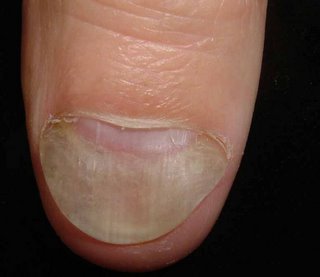
Nail Dystrophy

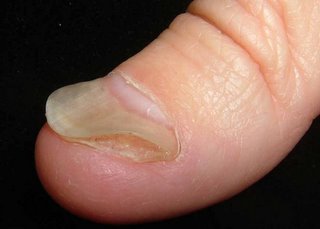
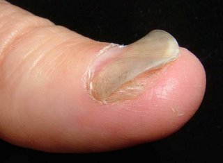
This 50 yo man has a three year history of a nail dystrophy.
I asked him to write out a detailed history:

"Started 18 months ago. Might well have contracted the condition while handling and cutting blocks of cheese as part of my former job. I used to handle and work with dozens of varieties of often moldy cheeses for three hours per week.

The condition began with a raised red ridge near the thumbnail. It was a connected series of raised red lumps about 1 to 2cm long and 3 or 4mm wide. This condition traveled progressively up and around my thumb. It always progressed one way up my thumb with the old area healing. It became a migrating „"ing of red". This went on for about six months. Often complete healing would occur, but then it would come up again right where it had left off and progress some distance more. Eventually it completely died out a little ways into the palm. There has been no trace of this symptom for a year now.

But starting also about 18 months ago the thumbnail became discolored (white and yellowish) and detached from the skin underneath. I've cut the thumbnail all the way back to the base in the hopes it will grow out correctly, but it remains detached from the skin underneath. Also the nail is misshapen: it is raised in some places and sunken in the middle. This has been the growth pattern for more than a year now. The skin underneath is clean but has a slight coating of what seems to be nail tissue on it, thus making it harder for the nail to attach itself. The nail is sound, just wavy and not attached underneath; because it is unattached underneath it is not pink, but rather it is white.


In the dermatology office there have been 2 negative KOH preps and a negative fungal culture. I did see some hyphae on one occasion. Ths question is: Is this a variant of funal nail disease, or are we dealing with onycholysis alone.

Your thoughts are appreciated.