This is default featured slide 1 title

Go to Blogger edit html and find these sentences.Now replace these sentences with your own descriptions.

This is default featured slide 2 title

Go to Blogger edit html and find these sentences.Now replace these sentences with your own descriptions.

This is default featured slide 3 title

Go to Blogger edit html and find these sentences.Now replace these sentences with your own descriptions.

This is default featured slide 4 title

Go to Blogger edit html and find these sentences.Now replace these sentences with your own descriptions.

This is default featured slide 5 title

Go to Blogger edit html and find these sentences.Now replace these sentences with your own descriptions.

Showing posts with label lichen planus. Show all posts

Friday, October 4, 2013

Nonspecific Oral and Genital Lesions

Abstract:  92 year-old woman with 6 year history of painful oral and genital lesions

HPI:  The patient is an otherwise healthy and alert 92 year old woman with painful erosions of tongue and vulva for six years. She has marked pain with eating and urination.  Mouth lesions preceded genital lesions by a few years.  She has been followed for erosive lichen planus but it is unclear if any biopsies were positive.  She has lost weight because her painful tongue causes her to avoid eating.  She has been treated with clobetasol ointment,  Viscous Lidocaine was not effective.

O/E:  Erosive lesions of tongue and labia.  No bullae noted. Remainder of cutaneous exam unremarkable.

Clinical Photos:


Pathology:  Biopsy of genital labial skin shows papillary dermal fiborsis and a mild superficial perivascular mixed inflammatory cell infiltrate.  No lichenoid infiltrate.  Direct immunofluorescence from perilesional skin was negative.  PASD negative for fungi.

Diagnosis:  Painful erosions mouth and genitalia.  At present no support for lichen planus or bullous process histologically.

Questions:  What are your thoughts? Her oral pain makes eating difficult and negatively affects her quality of life.  Are there other therapies that we could consider?

Saturday, December 15, 2012

Lichen Nitidus

Presented by Yoon Cohen, D.O.

Abstract: 13 year-old boy with a few years history of lichen nitidus

HPI:  This is a 13 year-old Hispanic boy who presented for treatment of warts. During the exam, multiple small white shiny dots were noticed. The patient has been aware of these asymptomatic lesions for the past few years.  He had been given triamcinolone 0.1% ointment bid, but forgot to apply it because it did not bother him. 

O/E:  There  are multiple pinhead-sized 1 mm white shiny flat-topped papules in an oval shape on the right knee.  

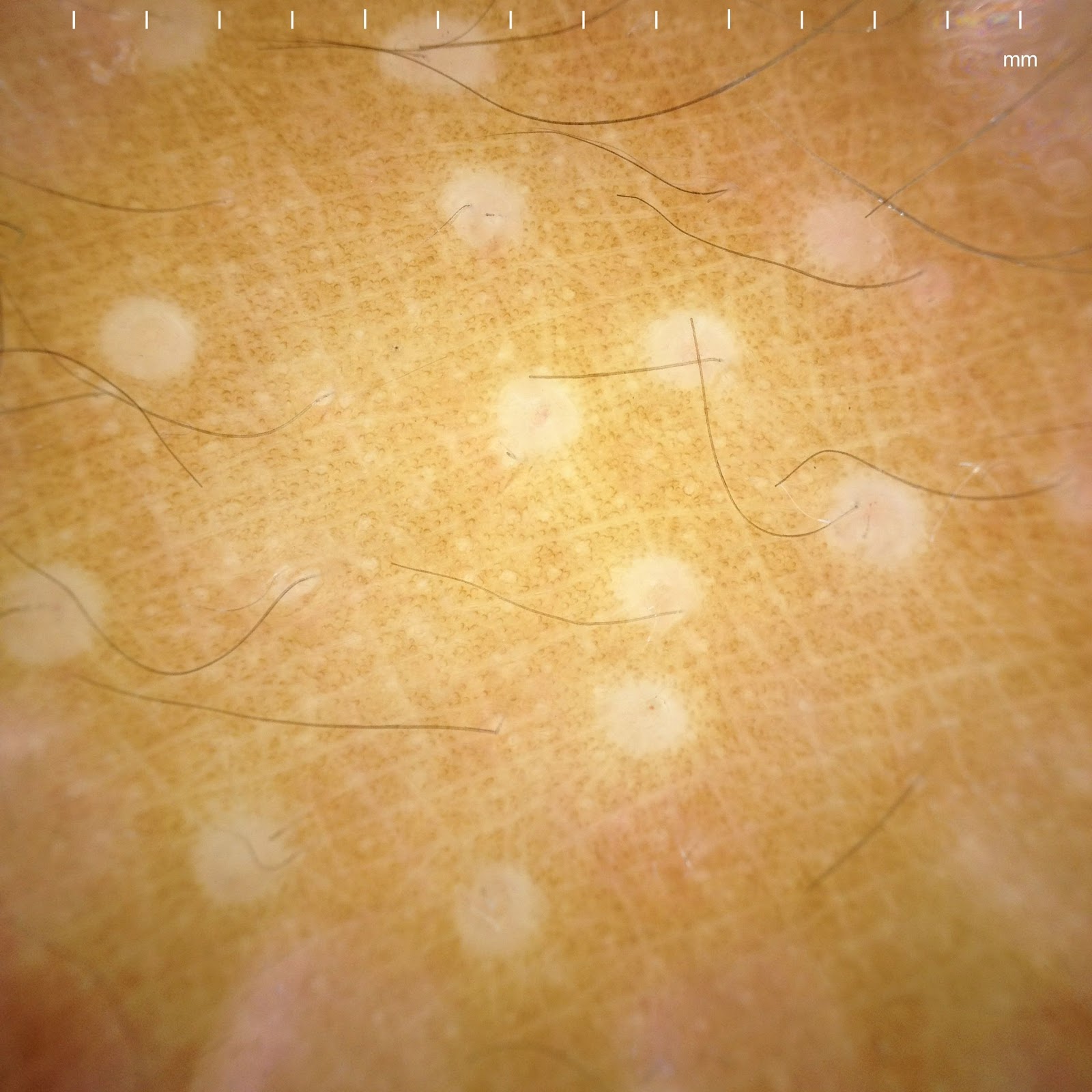
Dermatoscopic Photo:


Multiple small white circles
The dermoscopic image taken with the Canfield Dermatoscope with iPhone attachment

Diagnosis:

Lichen nitidus

Discussion:
While there are many dermoscopic studies on the skin cancers, there is a growing interest of dermoscopic features on other skin diseases such as inflammatory diseases, connective tissue disorders, or even psychogenic related skin diseases. Lichen nitidus is easily recognized clinically with experienced eyes. With my own interest in dermoscopy, I report an interesting dermoscopic finding of lichen nitidus. There are 1-2 mm multiple small white circles which represent subepidermal infiltrate of lymphocytes, histiocytes, and multinucleated giant cells in the dermal papillae. 

Lichen nitidus was first described by Pinkus in 1907. Lichen nitidus, which means shiny papules, is a relatively uncommon, asymptomatic, chronic eruption, consisting of minute sharply demarcated skin-colored papules. It has a predilection for males (4:1) and the mean age of onset was 7 yrs for males and 13 yrs for females. Typically, patients present for assessment with an asymptomatic or mildly pruritic eruption. The eruption may be localized to one or more areas or generalized in distribution. The pathogenesis of lichen nitidus is unknown. It was originally believed to be a form of tuberculid and more recently considered to be a variant of lichen planus. Histologially, lichen nitidus is characterized by a circumscribed collection of inflammatory cells in the papillary dermis that abuts the overlying epidermis. The inflammatory infiltrate consists of lymphocytes, histiocytes, and multinucleated giant cells. DIF negative.  Most cases require no treatment due to the asymptomatic nature of the eruption and tendency for spontaneous resolution. For symptomatic cases, moderately potent or potent topical steroid therapy may help. There are a number of anecdotal reports of improvement or clearance with narrow band UVB phototherapy, extensive sunlight exposure, oral astemizole, oral cetirizine-levamisol combination, topical dinitrochlorobenzene, itraconazole and oral cyclosporin. Oral retinoids have been used successfully in the treatment of palmoplantar lichen nitidus.

Reference:
Schachner LA, Hansen RC. Pediatric Dermatology. 4th edition. 2011
L. Nitidus eMedicine (free open access)

Comment: By DJ Elpern.  This is the first report of the dermatoscopic appearance of L. nitidus.  Yoon Cohen deserves credit for this observation.