This is default featured slide 1 title

Go to Blogger edit html and find these sentences.Now replace these sentences with your own descriptions.

This is default featured slide 2 title

Go to Blogger edit html and find these sentences.Now replace these sentences with your own descriptions.

This is default featured slide 3 title

Go to Blogger edit html and find these sentences.Now replace these sentences with your own descriptions.

This is default featured slide 4 title

Go to Blogger edit html and find these sentences.Now replace these sentences with your own descriptions.

This is default featured slide 5 title

Go to Blogger edit html and find these sentences.Now replace these sentences with your own descriptions.

Thursday, September 20, 2012

Incidentaloma

The patient is an 82 man who presented  for evaluation of a facial lesion.  That lesion was a basal cell carcinoma.  However a complete skin exam revealed a suspicious tumor on the right upper back. 

The patient has type III skin.  This "new" lesion on the back is an eight mm in diameter hypopigmented papule with an eccentrically placed  2 mm pigmented papule.

Clinical Photos:


Polarized View

Non-Polarized View


The Power of BLINK:
B = not benign
L = Lonely  1 point
I = Irregular pigment 1 point
N = (pt. unaware of lesion)
K = Known Dermatoscopic abnormalities  1 point
Score = 3  (Mandates biopsy)

The patient is scheduled for an excisional biopsy of the back lesion.  What are your thoughts at this time?

Biopsy shows this to be Malignant Melanoma
Type: unclassified
Thickness: 2.80 mm
Margins: signs of regression at ;ateral margin
Ulceration: absent
Mitoses: > 1/ mm squared
Vascular invasion: absent
Precursor lesion absent


Friday, September 14, 2012

Unusual Pigmentary Process

Presented by Dr. Ana Brasileiro
Central Lisbon Central Hospital
Lisbon, Portugal

Abstract:  51 yo man with 6 year history of pigmentary disorder.

HPI: The patient is a 51 year old male, born in Brazil and living in Portugal for 5 years. He reports that for the last 6 years he has experienced a slowly progressive dermatosis, first affecting the posterior cervical region and the superior part of the trunk, with progressive involvement of all the trunk, arms and legs. The patient denies pruritus or any other symptom. He has no contributory medical past story, and his only medication is occasional omeprazole 20mg. He has no relevant familiar history (neither the parents, brothers or children have any dermatological condition).

O/E:  This shows reticular pattern with hyper and hypopigmentation on the trunk extending to the arms and legs.

Clinical Photos:


Lab: Macrocytic anemia (Hb 11.5 g/L, VGM 106 fL) with normal levels of folic acid, B12 vitamin, as well as normal protein electrophoresis. Iron levels, ferritin and transferin are also normal (although these are not so relevant for a macrocytic anemia) The liver and kidney functions are within the normal range, and the tests for HIV, HCV and syphilis were negative. He has acquired immunity to HBV.

Histopathology:
The biopsy showed poikiloderma with amyloid deposits in the superficial dermis, consistent with cutaneous discromic amyloidosis.


Diagnosis: Amyloidosis cutis dyschromica

Questions: What is your opinion about this case?
From your experience, do you know of any case of dyscromic cutaneous amyloidosis starting at the age of 45?
Should we consider other diagnosis?
Do you think that the macrocytic anemia is somehow related with this cutaneous condition?

References: 

1. Amyloidosis cutis dyschromica in two female siblings: cases report; Yang et al. BMC Dermatology 2011, 11:4 “This disorder is characterized by the following features: (i) dotted, reticular hyperpigmentation with hypopigmented spots without papulation almost all over the body; (ii) no or little itchy sensation; (iii) onset before puberty; and (iv) small foci of amyloid closely under the epidermis “

2.Familial amyloidosis cutis dyschromica; Karadag, A.; Simsek, G; Turk J Med Sci 2010; 40 (1): 151-154 “In the literature, there are a few reports of familial cases (…). Cases accompanying autoimmune connective tissue diseases and primary biliary cirrhosis were reported in the other primary cutaneous localized amyloid types, such as macular and lichenoid amyloidosis (7). However, no another systemic and dermatological disease accompany ACD. In the literature there is only one case accompanying generalized morphea (5).”


Friday, September 7, 2012

Hidrotic Ectodermal Dysplasia

Abstract: 44 yo woman with Clouston Syndrome and severe painful kereatoderma

HPI: This 44 year-old disabled woman has the severe stigmata of Clouston Syndrome (CS-HED) (a variant of hidrotic ectodermal dysplasia seen in French Canadian probands).  She has impressive keratoderma of the feet which  make ambulating difficult and painful.  Her father had CS-HED as do three of her daughters.  The presentation in her daughters is variable.  Over the years, her keratoderma has become progressively severe. On a recent visit she said, "Please cut my feet off and feed them to the pigs."  She has been hospitalized in the past for cellulitis secondary to breakdown of plantar keratoderma.  The patient had seen a podiatrist who debrided her feet in theater on occasion, but she moved to another state and her insurance does not cover her there.

O/E:  This is a sad looking 44 year-old woman.  She has alopecia universalis, is edentulous, her nails are dystrophic and she has has abnormal and hyperkeratotic palms.  The most striking feature is massive keratoderma plantaris which tends to break down and become infected.

Clinical Photographs (presented with the patient's consent):


Diagnosis and Questions:   This unfortunate woman has massive keratoderma in the setting of Clouston Hidrotic Ectodermal Dysplasia.  Her painful feet make ambulating difficult and intolerable.  This has become more severe over the years and the patient has become discouraged.  She has asked on more than one occasion to have her feet amputated.  My plan at this point is to find a podiatrist or plastic surgeon who can do shave excisions to debride the keratoderma while she is on acitretin which will hopefully forestall the recurrence of her keratoderma.  She had a tubal ligation 22 years ago and is not sexually active at present but will be monitored for pregnancy monthly.  If this approach fails, deep excision of keratoderma with grafting has occasionally been reported.  I plan to start her on 10 mg of acitretin three times a week and increase slowly as tolerated.  
Your suggestions will be appreciated.

Tuesday, July 31, 2012

65 yo woman with nodulo-ulcerative leg lesions

Abstract:  65 yo woman with 4 year history of papules and nodules and 2 - 3 month history of papulo-ulcerative lesions on the legs

HPI:  This 65 yo woman has had leg lesions for ~ four years.  Initially, papules and small nodules. Over the past few months a number of these have become ulcerative.  She had lymphadenopathy two years ago and a lymph node biopsy was interpreted as sarcoidosis.  Her CXR was normal and no treatment was rendered,

O/E:  Healthy appearing woman with numerous 8 - 10 mm nodules on both lower extermities.  Similar lesions were ulcerative. Remainder of cutansous examination unremarkable.

Clinical Photos:


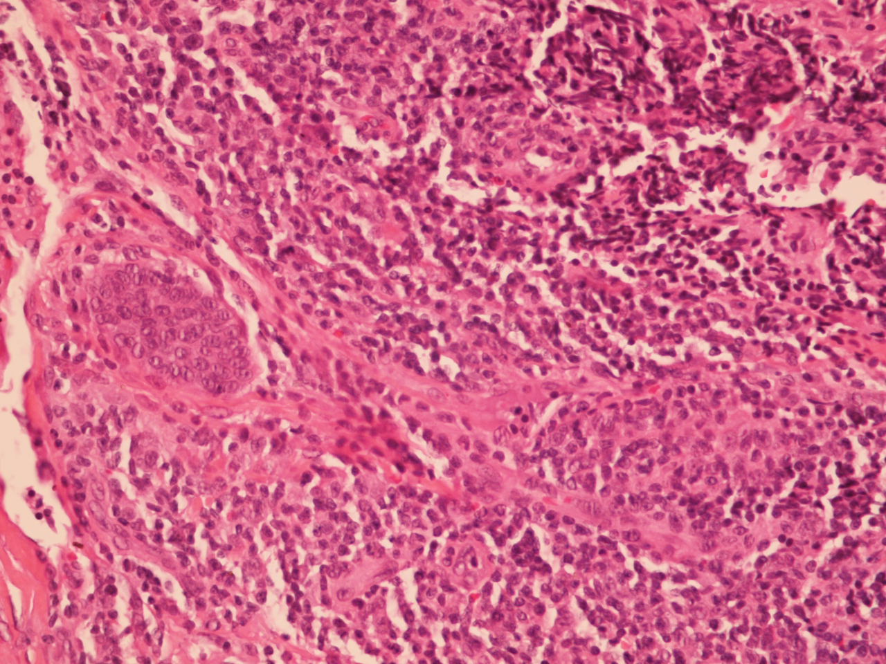
Pathology:  Small noncaseating epitheliod granulomas containing multinucleated giant cells within a dense fibrotic stroma and a perivascular plasmacytic infiltrate at the periphery.  Rare elongate acid-fast bacilli are seen in two granulomas.
4x

10x
 
20x
 
40x (AFB)


Diagnosis: Necrobiosis-like variant of sarcoidosis.  What is the significance of rare AFBs?  She will have PPD and work-up to rule out TBC.

Questions:  Do you have alternative diagnoses? How would you treat her?


References:
1.  Necrobiosis lipoidica-like skin lesions in systemic sarcoidosis.
Igawa K, Maruyama R, Satoh T, Yokozeki H, Katayama I, Nishioka K.

J Dermatol. 1998 Oct;25(10):653-6.
Abstract
A 62-year-old woman with systemic sarcoidosis developed erythematous plaques on her lower legs. Clinically, two kinds of skin lesions were distinguished; one type formed brownish-red plaques with induration suggesting plaque-type skin sarcoid, and the other formed purplish erythematous plaques with atrophic centers resembling necrobiosis lipoidica. In spite of this clinical appearance, a biopsy specimen from one of the latter lesions revealed typical skin sarcoid histology composed of discrete non-caseating granulomas, while that from one of the other lesions showed necrobiotic changes of collagen bundles surrounded by epitheloid histiocytes and foreign-body giant cells. Because cutaneous involvement of sarcoidosis may mimic necrobiosis lipoidica clinically and/or histologically, we diagnosed her skin lesions as necrobiosis-like skin sarcoid.

2. Histologic observations of variably acid-fast pleomorphic bacteria in systemic sarcoidosis: a report of 3 cases.
Cantwell AR Jr. Growth. 1982 Summer;46(2):113-25.
Abstract: Tissue sections of skin and lymph nodes from three consecutively diagnosed cases of systemic sarcoidosis were studied for the presence of acid-fast bacteria, utilizing routine and acid-fast staining techniques recently recommended for the demonstration of cell-wall-deficient bacteria (L-forms). Evidence of variably acid-fast cocco-bacillary forms was present within the biopsy material of all the patients. The combined findings of variably sized, predominantly coccoid forms, along with larger forms resembling L-form "large bodies," and short acid-fast rods all suggest that cell-wall-deficient bacteria (possibly related to the mycobacteria or corynebacteria) may be present in cases of sarcoidosis. 

Monday, July 23, 2012

Deadline Looming

Abstract:  27 year-old woman with severe generalized contact dermatitis secondary to poison ivy, starting two weeks before her wedding.

HPI:   This 27 yo woman, whose wedding is August 4th, 2012, presented on July 19th with a one day history of a facial eruption.  She has a history of rhus allergy and had been at the beach collecting leaves for an architecture project a couple of days before the onset of symptoms.  At the time, she was wearing a bathing suit and a wrap-around towel.   The rash first appeared on her face and left lower abdomen.  She has a history of acne vulgaris which is quiescent now.  She was on prednisone 20 and 10 mg a day for a week, ~ 10 days before this episode.

O/E:  July 19th:  Marked erythema and induration on left forehead, cheek, chin.  Vesicles and small bullae scattered in the area;  A few erythematous streaky patches lsft hip.  Over the next few days, in spite of treatment the process progressed to involve abdomen, neck and fingers.  On July 23, there are new erythematous-bullous areas on the left lower abdomen and hip. (see photos).

Clinical Photos:  See below

Therapy: 
July 19th:  Prednisone 20 mg b.i.d. (her weight is 54 kg).  Cool tap water compresses.  Hydroxizine 20 - 30 mg hs.
July 21:  Because of progression of dermatitis, prednisone increased to 50 mg per day in divided doses.  Dome Boro compresses 3 x per day, Silvadene cream because of some erosions at site of bullae.  Prednisone is causing her to feel anxious and panicky.  Lunesta 1 - 2 mg added for sleep. 
July 23:  I am surprised that new lesions continue to develop in spite of an adequate dose of prednisone (see photos).  Have added a "soak and smear" protocol for body lesions and desoximetasone cream bid for dermatitis on body and once daily for face.

Diagnosis: Severe Allergic Phyto-Contact Dermatitis secondary to rhus.  We saw this at onset and in spite of a reasonable dose of prednisone and cool compresses it has progressed.  Steroid dose was limited because of CNS symptoms and initially I was reluctant to use topical steroids due to her history of acne; but have just started desoximetasone cream on 7/23.  The timeline is important as she is getting married in 12 days.  It is odd that this has progressed after what is usually an adequate dose of prednisone, and I am worried that increasing the prednisone may cause more anxiety and insomnia. Most likely this is a severe anamnestic response to urushiol and she may indeed need a higher dose of prednisone or she may not be absorbing it.

Questions:
1) How would you handle the facial erythema?

2) Topical corticosteroids as well as oral steroids can exacerbate acne.  Should we add a moderate to strong topical corticosteroid for a few days to suppress erythema?

3) She is anxious and has insomnia secondary to prednisone already.  I feel prednisoen is the key to improvement, but am reluctant to push the dose.  Your thoughts?

4) Is there a role for topical tacrolimus?

Your suggestions re: diagnosis and management will be appreciated.

Photos:
July 19


July 20

July 21

July 22

July 23
New lesions 4 d p start prednisone

July 24
Real Improvement Noted Today

7/30
Pretty Much Better

Tuesday, July 10, 2012

Nasal Tumor for Diagnosis

Presented by Henry Foong, Ipoh, Malaysia


Abstract: 45 yo man with one year history of a tumor on the nasal tip.

HPI: The patient is a 45 yr old man with a growth on the tip of nose for a year. It started as an asymptomatic papule last year and was shaved off by a GP.  He remained well till 3 months ago when it started to recur.  He had seen few doctors and the last doctor, a plastic surgeon decided to refer to a dermatologist. He lives in Teluk Intan about 100 km south from Ipoh. He is an excavator driver, does mainly outdoor work and has no family history of skin cancer.

O/E: Examination showed a firm non tender nodule 1.5 x 1.5 cm on the tip of the nose with a crateriform centre. Regional nodes were not enlarged.  

Clinical Photos:


Pathology: An incisional biopsy was done on the tumor. Biopsy done showed diffuse and nodular infiltration of lymphocytes in the reticular dermis and deeper layers, separated by variable amount of fibrocollagenous tissue. Admixed with the lymphocytes are few eosinophils and plasma cells. There is no epidermotropism. The epidermis is unremarkable. Mitotic figures are discernable.  

Immunohistochemical studies were performed on the tissues. The lymphoid population showed a polyclonal population of both T cell and B cell markers. A diffuse positivity of pan T markers CD 3 along with CD5 were noted. Patchy distribution of the B cell markers throughout is noticed for CD20, CD79a, PAX5 and MUM 1. Ki67 shows a proliferation index of 30-40% in the entire population.


Diagnosis (Tentative): Lymphocytoma Cutis

Questions:  Is the immunohistochemistry supportive of lymphocytoma cutis (cutaneous B-cell pseudolymphoma)  or cutaneous lymphoma? Will excision of the tumor help? If surgery is not contemplated, what would be recommmended treatment?


Sunday, July 1, 2012

Nail Dystrophy in a Nurse

A colleague requested our opinion regarding a woman who contacted him, but has not seen.  Here are her words and photos.
" I am a 61 yo palliative care nurse who has had a nail dystrophy for around 14 months. There is no pain or discomfort. I take no meds by mouth and am in good general health. Every 2 weeks I get a professional manicure and have never had acrylic, gel or silk.  I tried "formaldehyde free" products for a while, as well as Biotin 5000mcg daily and noted no improvement.  As part of my job, I visit patients in their homes and wash my hands before & after each visit. Rarely do I have the need to wear gloves.  Your suggestions will be welcome."


Questions at this time:
1) Are all nails involved (including toe-nails)?
2) Has a dermatologist been consulted and have KOH prep and/or fungal culture been done?