This 23 yo woman has had facial erythema and acuminate micropapular lesions since childhood. She has extensive keratosis pilaris of her arms and thighs in addition. She has not gone out in public without thick make-up for 10 years. Her fiance has never seen her without make-up. She is desperate to haved this treated. I suspect this is a variant of KP rubra facei, possibly with ulerythema oopryogenes

This is default featured slide 1 title
Go to Blogger edit html and find these sentences.Now replace these sentences with your own descriptions.

This is default featured slide 2 title
Go to Blogger edit html and find these sentences.Now replace these sentences with your own descriptions.

This is default featured slide 3 title
Go to Blogger edit html and find these sentences.Now replace these sentences with your own descriptions.

This is default featured slide 4 title
Go to Blogger edit html and find these sentences.Now replace these sentences with your own descriptions.

This is default featured slide 5 title
Go to Blogger edit html and find these sentences.Now replace these sentences with your own descriptions.
Tuesday, May 24, 2005


Saturday, May 7, 2005
Seven Year Old Boy with Chronic Dermatitis
A.D. is a 7 year-old boy who was adopted from Siberia by a family in western Massachusetts at 21 months of age. Since adoption he has had recurrent dermatitis on torso and extremities. The lesions are mostly nummular by history. They have responded to systemic antibiotics on occasion. He was seen here yesterday for the first time for a second opinion.
The child appears normal otherwise. He has no evidence of atopy. This is the largest lesion. All are plaques, all covered with some crust, intensely pruritic. I applied some pressure to the large plaque with a cotton tipped applicator and a small amount of creamy pus was extruded.
My working diagnosis is nummular eczema driven by hypersensitivity to staph. A culture was taken and I'll wait for results before treating. This has been going on for 5 years. I may do a biopsy, but am not sure it will be helpful.
Any thoughts would be appreciated. The parents are at their wits end. He has also been treated with mupirocin cream in past with some success. Tacrolimus was not helpful.
The culture grew out coagulase positive staph sensitive to everything; even Penicillin G. This is unusual in the U.S. where most Saph is resistant to penicillin. One wonders if this is a strain he brought over from Russia when he was adopted. I started him on Pen VK 250 mg qid. I will add a topical corticosteroid and mupirocin - the latter for nares and crural folds. Will give follow-up after a couple of weeks. If he continues to have staph infections like this, I will look into his Ig status.
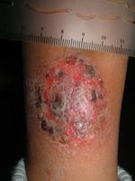
Right leg
The child appears normal otherwise. He has no evidence of atopy. This is the largest lesion. All are plaques, all covered with some crust, intensely pruritic. I applied some pressure to the large plaque with a cotton tipped applicator and a small amount of creamy pus was extruded.
My working diagnosis is nummular eczema driven by hypersensitivity to staph. A culture was taken and I'll wait for results before treating. This has been going on for 5 years. I may do a biopsy, but am not sure it will be helpful.
Any thoughts would be appreciated. The parents are at their wits end. He has also been treated with mupirocin cream in past with some success. Tacrolimus was not helpful.
The culture grew out coagulase positive staph sensitive to everything; even Penicillin G. This is unusual in the U.S. where most Saph is resistant to penicillin. One wonders if this is a strain he brought over from Russia when he was adopted. I started him on Pen VK 250 mg qid. I will add a topical corticosteroid and mupirocin - the latter for nares and crural folds. Will give follow-up after a couple of weeks. If he continues to have staph infections like this, I will look into his Ig status.

Right leg

Tuesday, April 26, 2005
A Change of Pace
The patient accompanied her "mother" to the office. She is a 12 yo chihuaha with autoimmune thyroid disease and extensive alopecia areata. I was aksed what could be done and injected large areas of her skin with intralesional triamcinalone. Unfortunately, there is no billing code for this - the dog lacked health insurance.
(Actually, I deferred treatment until I hear from my august colleagues)
Note tail alopecia in Fig. 3

Fig 1
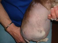
Fig 2
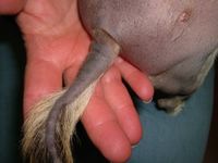
Fig. 3
Ref:
A natural canine homologue of alopecia areata in humans.
Tobin DJ, Gardner SH, Luther PB, Dunston SM, Lindsey NJ, Olivry T.
Department of Biomedical Sciences, University of Bradford, Bradford, UK.
Br J Dermatol. 2003 Nov;149(5):938-50.
BACKGROUND: Alopecia areata (AA) is suspected to be an autoimmune disease directed preferentially against hair follicles (HF) affecting both humans and various mammalian species. Recently, two rodent models of AA were described, namely the ageing C3H/HeJ mouse and the DEBR rat. Despite several case reports of canine AA in the literature, there has been no systematic assessment of the disease in these companion animals, and it is also not known whether dogs with AA could be useful as an outbred homologue of this disease in humans. OBJECTIVES: To evaluate the clinical, histopathological and immunopathological features of 25 dogs with AA and compare these data with those found in the human disease. PATIENTS/METHODS: Twenty-five client-owned dogs exhibiting macroscopic alopecia with peri- or intrabulbar lymphocytic infiltrates were selected for study. Biopsies and sera were obtained and assessed by histopathology, direct immunofluorescence of immunoreactant deposition, immunohistochemistry for lymphocyte markers, indirect immunofluorescence and immunoblotting analysis of circulating serum IgG, selective immunoprecipitation of HF proteins by serum IgG, and passive transfer of purified canine IgG into naive C57BL/10 mice. RESULTS: Clinical signs including alopecia, skin hyperpigmentation and leucotrichia usually developed during adulthood and were first seen on the face, followed by the forehead, ears and legs. Spontaneous remission of alopecia occurred in 60% of dogs and regrowing hair shafts were often non-pigmented. Histological examination of skin biopsy specimens revealed peri- and intrabulbar mononuclear cell infiltrates affecting almost exclusively anagen HF. Direct immunofluorescence analysis detected HF-specific IgG in 73% of dogs, while indirect immunofluorescence revealed circulating IgG autoantibodies to the HF inner and outer root sheaths, matrix and precortex. Immunoblotting analysis revealed IgG reactivity to proteins in the 45-60 kDa molecular weight range and with a 200-220 kDa doublet. The latter was identified as trichohyalin by selective immunoprecipitation. Purified HF-reactive IgG, pooled from AA-affected dogs, was injected intradermally to the anagen skin of naive mice where it was associated with the local retention of HFs in an extended telogen phase in AA-treated skin compared with that seen in controls. CONCLUSIONS: These findings are very similar to those reported for human AA patients; therefore, they support the consideration of dogs with AA as a useful homologue for the study of the pathogenesis of this common autoimmune disease of humans.
(Actually, I deferred treatment until I hear from my august colleagues)
Note tail alopecia in Fig. 3

Fig 1

Fig 2

Fig. 3
Ref:
A natural canine homologue of alopecia areata in humans.
Tobin DJ, Gardner SH, Luther PB, Dunston SM, Lindsey NJ, Olivry T.
Department of Biomedical Sciences, University of Bradford, Bradford, UK.
Br J Dermatol. 2003 Nov;149(5):938-50.
BACKGROUND: Alopecia areata (AA) is suspected to be an autoimmune disease directed preferentially against hair follicles (HF) affecting both humans and various mammalian species. Recently, two rodent models of AA were described, namely the ageing C3H/HeJ mouse and the DEBR rat. Despite several case reports of canine AA in the literature, there has been no systematic assessment of the disease in these companion animals, and it is also not known whether dogs with AA could be useful as an outbred homologue of this disease in humans. OBJECTIVES: To evaluate the clinical, histopathological and immunopathological features of 25 dogs with AA and compare these data with those found in the human disease. PATIENTS/METHODS: Twenty-five client-owned dogs exhibiting macroscopic alopecia with peri- or intrabulbar lymphocytic infiltrates were selected for study. Biopsies and sera were obtained and assessed by histopathology, direct immunofluorescence of immunoreactant deposition, immunohistochemistry for lymphocyte markers, indirect immunofluorescence and immunoblotting analysis of circulating serum IgG, selective immunoprecipitation of HF proteins by serum IgG, and passive transfer of purified canine IgG into naive C57BL/10 mice. RESULTS: Clinical signs including alopecia, skin hyperpigmentation and leucotrichia usually developed during adulthood and were first seen on the face, followed by the forehead, ears and legs. Spontaneous remission of alopecia occurred in 60% of dogs and regrowing hair shafts were often non-pigmented. Histological examination of skin biopsy specimens revealed peri- and intrabulbar mononuclear cell infiltrates affecting almost exclusively anagen HF. Direct immunofluorescence analysis detected HF-specific IgG in 73% of dogs, while indirect immunofluorescence revealed circulating IgG autoantibodies to the HF inner and outer root sheaths, matrix and precortex. Immunoblotting analysis revealed IgG reactivity to proteins in the 45-60 kDa molecular weight range and with a 200-220 kDa doublet. The latter was identified as trichohyalin by selective immunoprecipitation. Purified HF-reactive IgG, pooled from AA-affected dogs, was injected intradermally to the anagen skin of naive mice where it was associated with the local retention of HFs in an extended telogen phase in AA-treated skin compared with that seen in controls. CONCLUSIONS: These findings are very similar to those reported for human AA patients; therefore, they support the consideration of dogs with AA as a useful homologue for the study of the pathogenesis of this common autoimmune disease of humans.
Sunday, April 24, 2005
Asymptomatic Blanching Palmar Eruption
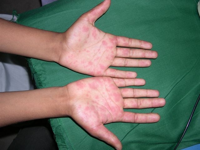
Asymptomatic non blanching eruption

A 14-year-old, otherwise healthy boy presented with an asymptomatic red blanching eruption on the palms that developed about one week earlier. Soles were not involved. No inciting factor including drugs or illness could be recognized. Rest of the physical examination and routine blood and urine tests were unremarkable. What conditions can give rise to this eruption and how should we work it up ?
Shahbaz A Janjua MD
Saturday, April 23, 2005
Case for diagnosis (Alberta, Canada)
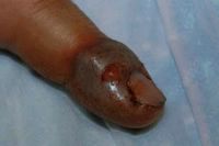
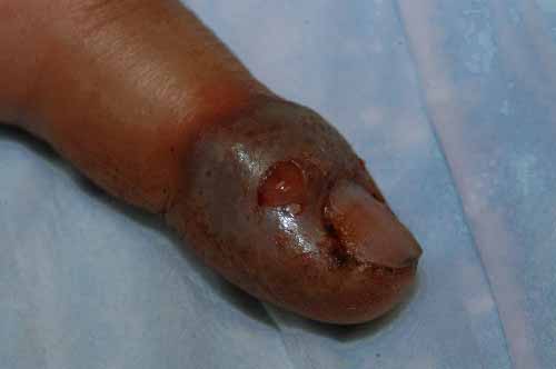
Day #1 this guy helped deliver a breech calf by c-section which was so traumatic that the calf was still-born and then the following day he helped butcher a cow. On Day #3 he noted swelling of his left index finger and by
Day #4 it blew up to look like these pictures. The more I read about orf the more I feel that this is likely the case. The risk factors are there and the time of year and setting is classic. Anyway, what do the viewers think?
(The break in the skin/expelled fluid in the pictures is my doing. There
were no vesicles or absesses and the tissue was tough and non-fluctuent.)
Duncan

Day #4 it blew up to look like these pictures. The more I read about orf the more I feel that this is likely the case. The risk factors are there and the time of year and setting is classic. Anyway, what do the viewers think?
(The break in the skin/expelled fluid in the pictures is my doing. There
were no vesicles or absesses and the tissue was tough and non-fluctuent.)
Duncan

Sunday, March 27, 2005
20 yo man with cutaneous larva migran

The patient is a 20-year-old man who noticed multiple intensely pruritic creeping lesions on his abdominal wall for more than a month. He is a body building enthusiast.
Examination showed multiple 2-3mm wide serpiginous raised erythematous tunnels on the left periumbilical and right upper quadrant of the abdominal wall.
Treatment was with freezing 1cm distal to the end of the trail with liquid nitrogen and adding oral mebendazole (zentel) 200mg bd for 3 days. It was successfully eradicated within a week of treatment.
Henry
Wednesday, March 23, 2005
10 year old girl with endogenous cheilitis
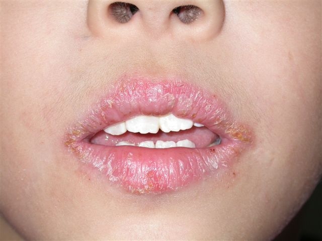
This patient presented with recurrent dry and fissured lips for more than 2 years. She has a strong family history of atopy. Her brothers and sisters have asthma and allergic rhinitis. Fortunately she didn't use any lipstick.
Examination showed fissures and dryness on both the upper and lower lips. There were crusts formations on the lips
Clinically she has endogenous (atopic) cheilitis
There are few factors that need to be considered here. is there a contact allergen element here? Lipstick would be the biggest culprit here. Is there a need to do a patch test?
Irritants can be an aggravating factor too. Lip smacking and hot spicy food can aggravate the eczema. Even toothpaste with strong mint flavour can be a factor too. I have seen several patients whose cheilitis was aggravated by our local "Darlie" toothpaste.
Moisturisers eg vaselin would be very helpful and they can be applied 4-5 times daily. Mild topical corticosteroids eg 1% hydrocortisone ointment or cutivate ointment helps. I prefer ointment to cream base in this situation.
Henry
Subscribe to:
Posts (Atom)


