This is default featured slide 1 title

Go to Blogger edit html and find these sentences.Now replace these sentences with your own descriptions.

This is default featured slide 2 title

Go to Blogger edit html and find these sentences.Now replace these sentences with your own descriptions.

This is default featured slide 3 title

Go to Blogger edit html and find these sentences.Now replace these sentences with your own descriptions.

This is default featured slide 4 title

Go to Blogger edit html and find these sentences.Now replace these sentences with your own descriptions.

This is default featured slide 5 title

Go to Blogger edit html and find these sentences.Now replace these sentences with your own descriptions.

Monday, October 31, 2005

Localized Hyperkeratosis

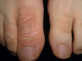
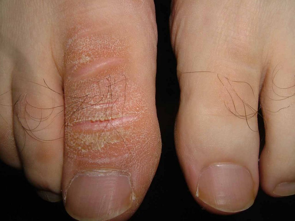
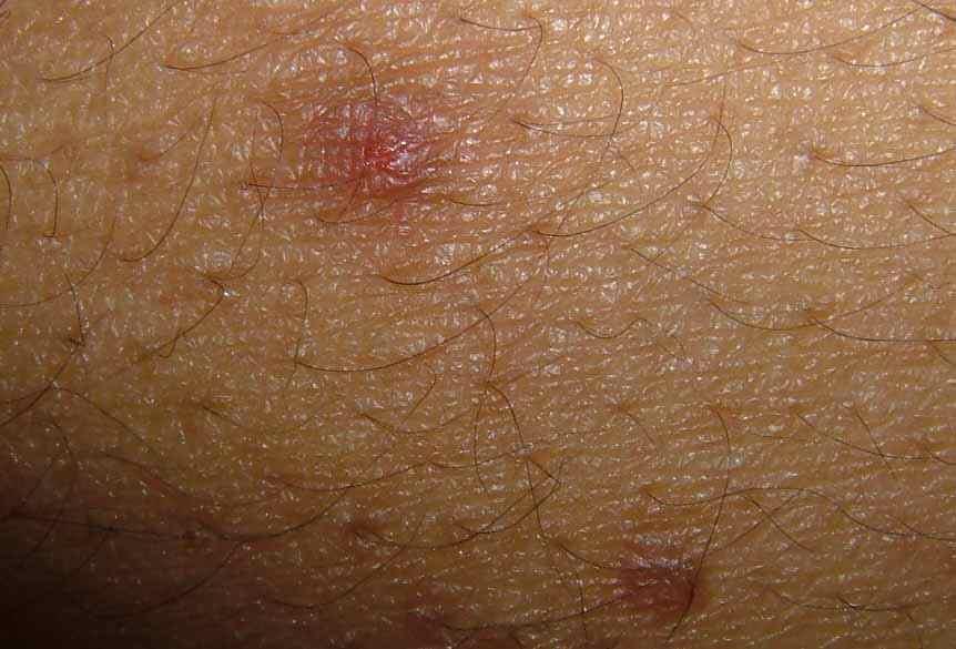
39 yo man with > 15 year hx of localized hyperkeratosis of right great toe. No unusual trauma to area. He's used mostly over the counter emollients without much help.

Are you aware of any localized forms of hyperkeratosis?

I have started him on Salex cream.

Pictured below are his affected right and normal left great toes.

Recent Onset Acne in Young Woman


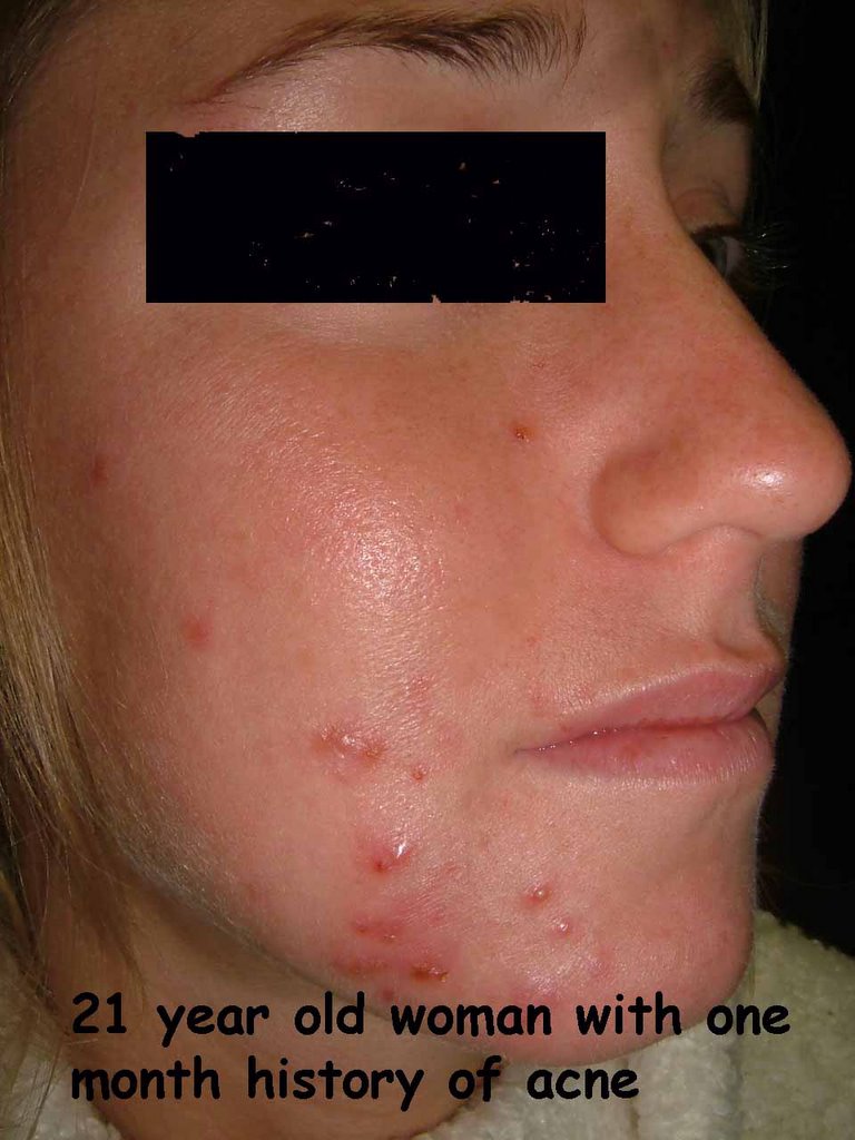
This 21 yo woman experienced the sudden onset of acne one month ago.
No new med. No unusual stresses.
She has never had acne like this before.

Do you think this is a mild case of pyoderma faciale rather than typical acne?

Sunday, October 30, 2005

BCC Scalp


The patient is a 75 yo man with a three month hisory of a lesion at the vertex of the scalp.
Exam: Man with Type II - III skin. 2 x 1.6 cm diameter erythematous plaque scalp. There are some small crusted areas.
Biopsy shows superficial BCC.

Question: Is Aldara appropriate?
C + E will take ages to heal.
Excision could n ot be closed without a graft or a flap.

Wednesday, October 26, 2005

19 year old student with acneiform eruption


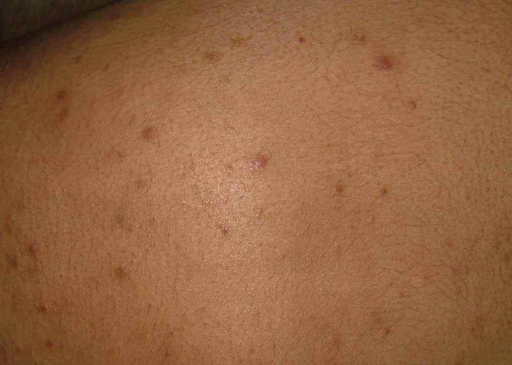
History: 19 year-old man with a 5 year history of acneiform eruption predominately on torso and proximal extremitiesl Some facial involvement, but less than on torso. He is in good health, no history of diabetes or steroid use. No antibiotics for months. In the past he has used benzoyl preoxide ceams and washes, topical retinoids, topical antibiotics and tetracycline and its derivatives. Mome has ever helped.

Exam: Healthy young man with Type IV skin. On torso and proximal extremities he has discrete erythematous papules and an occasional pustule. No cysts. Face largely clear.


Lab: nil

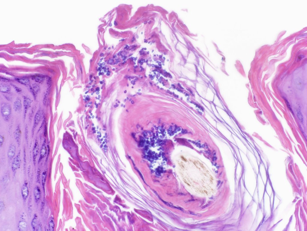
Pathology: Initial reading showed marked perifollicular lymphoneutrophilic infiltrate c/w acute folliculitis. PAS negative.


Diagnosis: Probable Pityrosporon folliculitis

Discussion: Are the yeasts incidental or indicative of pityrosporum folliculitis. I asked for more cuts and this showed numerous PAS (+)"fungal spores" in the follicular ostia.
I have started him on itraconazole 200 mg per day and Nizoral 2% shampoo to torso. The literature does not have good guidelins for how to treat this; but I suspect 4 - 6 weeks woth oral meds.
Please suggest diagnostic and therapeutic alternatives.

Thursday, October 20, 2005

Atypical Nail Pits


Case for Discussion:
This 40 year-old woman presented for evaluation of a nail dystrophy present for 2-3 months.
She has a history of Hashimoto's thyroiditis. About 9 months ago she developed vitiligo.
Her health is otherwise normal
Meds. Synthyroid and iron
Lab: Thyroid Perox AB 248 (Nl. 0 - 34 IU/ML)
ANA < 1:40

Physical Exam:
Vitiligenous patches left neck and left upper back
Around 4 finger nails show a distinctive pitting. The pits are fairly uniform and the affected nails are rough and lusterless. There are no cutansous lesions of psoriasis.

Discussion and Questikon:
The picture is atypical for psoriatic nail pits but that is not excluded.
I favor a relationship to the underlying autoimmunity that has caused the Hashimoto's and vitiligo.
The picture is similar to that seen with alopecia areata, but the patient has not has any alopecic patches.
Nail dystrophy has occasionally been described before the development of A. areata. And patients with Hashimoto's thyroiditis have a higher than expected incidence of alopecia areata.
We welcome your thoughts or suggestions.

Saturday, August 13, 2005

Hemangioma of Auricle


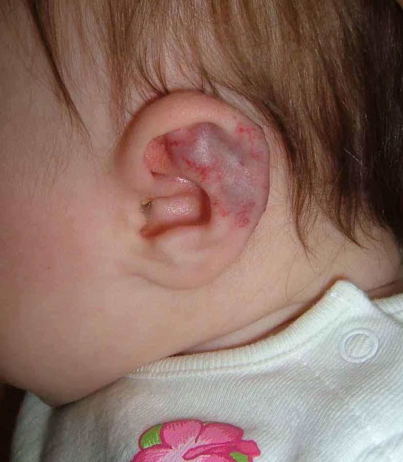
This 4 month infant girl was born with hemangiomas of the chin and left ear.
The lesion on the chin (not pictured) measures 1 cm in diameter and is typical of a congenital hemangioma.

The abnormality of the left ear involves the triangular fossa and the helix. I am concerned that as this involutes it could cause disfigurement. At the same time, I wonder if anyone has experience handling similar lesions in this site. A PubMed site found only a paucity of pertinent references.

Ref:
Cavernous hemangioma of the external ear canal.
Reeck JB, Yen TL, Szmit A, Cheung SW.
Laryngoscope. 2002 Oct;112(10):1750-2. Related Articles, Links
Division of Otology, Neurotology and Skull Base Surgery, Department of Otolaryngology-Head and Neck Surgery, Veterans Administration Medical Center, San Francisco, California, USA.

OBJECTIVE: To document the occurrence of a cavernous hemangioma of the external ear canal and to review the relevant literature.STUDY DESIGN Case report and literature review. METHODS: Review of a patient chart, imaging studies, operative report, and histologic findings. RESULTS: A cavernous hemangioma of the external ear canal not involving the tympanic membrane was surgically excised without complication. This is the third documented cavernous hemangioma of the external ear canal without tympanic membrane involvement in the English literature. Computed tomography scan is invaluable to narrow the differential diagnosis. Complete removal is curative. CONCLUSIONS: Cavernous hemangioma of the external ear canal with or without tympanic membrane involvement is a rare otologic entity amenable to surgical treatment. Temporal bone computed tomography scan imaging is an important preoperative diagnostic tool.

Tuesday, August 9, 2005

Nevus, Congenital


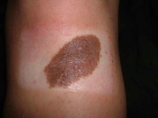
This 20 yo college student presented in consultation for removal of a congenital nevus.
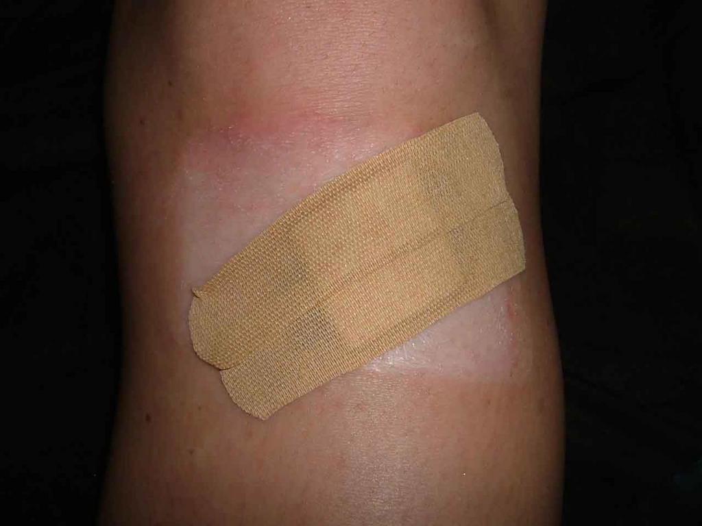
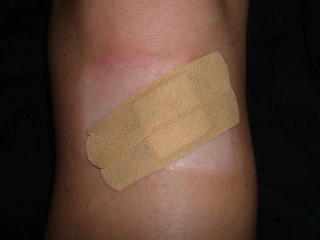
The lesion is located just distal to her left knee. She's been embarrassed about this since childhood and for the past six or seven years won't let anyone see it. Indeed she covers it at all times with bandaids.
The lesion measures 5.2 cm in diameter.
I have referred her to a surgeon for removal since I think that's better than psychotherapy. To minimize the scar it may need staged excision.
The young woman appears normal in all other respects - she is very upset about the lesion and the questions she gets about it.
I wonder if this has been reported. I would hardly call this Body Dysmprphic Syndrome.
Your thoughts are appreciatged.

Not much written about this area -- here's a ref that may touch on it:


Congenital melanocytic nevi. Evaluation and management.
Marghoob AA., Dermatol Clin. 2002 Oct;20(4):607-16, viii.
Department of Medicine, Dermatology Division, Memorial Sloan-Kettering Cancer Center, 1275 York Avenue, New York, NY 10021, USA. marghooa@mskcc.org

This article discusses the care of patients with CMN, who often require
a multidisciplinary approach involving pediatricians, family physicians,
internists, dermatologists, psychologists, plastic surgeons,
neurologists, and radiologists. The cosmetic and psychosocial issues,
combined with the knowledge of the increased risk of developing
melanoma or NCM, is a huge burden that many of these patients
and their families have to carry. This article describes the importance
for physicians to help these patients and families come to terms
with these issues, as well as remind their patients and their family
members that although melanoma, NCM, or other complications can
develop, most affected individuals do not develop any complications.
The article mentions that there are many healthy, happy, functional
adults with large, small, and multiple CMN alive today.