This is default featured slide 1 title

Go to Blogger edit html and find these sentences.Now replace these sentences with your own descriptions.

This is default featured slide 2 title

Go to Blogger edit html and find these sentences.Now replace these sentences with your own descriptions.

This is default featured slide 3 title

Go to Blogger edit html and find these sentences.Now replace these sentences with your own descriptions.

This is default featured slide 4 title

Go to Blogger edit html and find these sentences.Now replace these sentences with your own descriptions.

This is default featured slide 5 title

Go to Blogger edit html and find these sentences.Now replace these sentences with your own descriptions.

Wednesday, June 28, 2006

Eye Lash Alopecia

The patient is an eight year old girl who has had a dermatitis of her eyelid margins off an on for around a year. She gets crusted areas which itch and her mother feels she plucks her eyelashes. On the day of the visit the lids looked quiet, but there was definite loss of some eyelashes. There is some evidence of broken lashes.

Question:
Is this eyelash trichotillomania or isolated alopecia areata of the eyelashes? I favor trichotillomania
What is the prognosis for spontaneous recovery?

My plan is to see her if and when the dermatitis is more acute and obtain a culture to see if she has a bacterial blepharitis.


Saturday, June 24, 2006

Bed Bugs

This 35 year-old man went to Mamaronek, New York to watch the New York Open Golf Championship last weekend. The closest motel room he could find was in Bridgeport, Connecticut. The latter is a city with a fairly poor reputation. Two days after returning home to Massachusetts he developed the lesions pictured here on arms and to a lesser extend on torso and legs.

They are consistent with bedbug bites (Cimex lenticularis). For a brief description go to: http://www.emedicine.com/derm/topic600.htm

In November of 2005 a bedbug epidemic was reported in New York in the New York Times. I saw a couple of cases from there here in Williamstown, MA, 150 miles away. I suppose there's a similar problem in Bridgeport now.


Tuesday, June 20, 2006

Lyme Disease

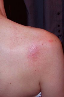
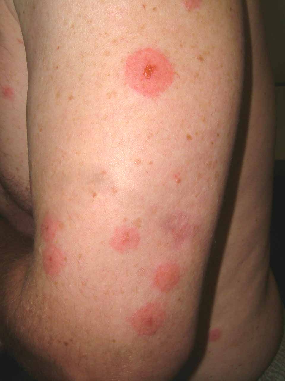
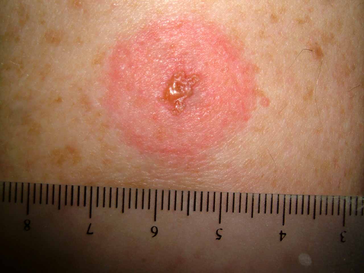
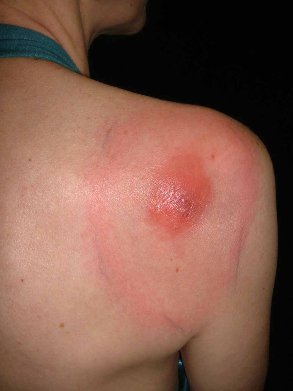
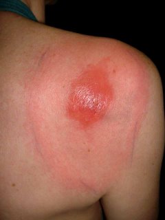
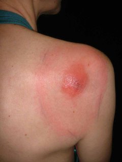
This 52 year-old woman presented as a walk-in patient today. She has a five day history of an erythematous nodule on the right shoulder which has developed a ring around it. The ring measures 9 by 11 centimeters in diameter. Initially, the nodule was painful for a few days; but the pain has subsided. She has had no fever or constutional symptoms and has continued to work as a school teacher.

The patient lives in rural New Hampshire and gardens a fair bit. She was seen two times at a primary care clinic where a diagnosis of spider bite was made. First she was given Keflex, then amoxicillin.

Diagnosis: I am uncertain. I favor Lyme Disease but this has atypical features. The central nodule is unusual, although the peripheral ring looks like Lyme. I also considered atypical Sweet's Disease and a pyogenic process. Lastly, I thought of eosinophilic cellulitis (Well's Syndrome) which I have never seen.

Plan: Since she feels well, I prescribed Doxycycline 100 mg bid, and ordered a cbc, Lyme titers and a G6PD (if the lesion becomes necrotic may want to try Dapsone. I considered biopsy but was strapped for time and thought Lyme most likely. Now, I am uncertain. Biopsy will be done when she is seen in follow-up unless she is significantly better.

Your thoughts will be welcome.


Addendum:

Path Report:
DIAGNOSIS: Skin - (A) Right Shoulder-Periphery-Ring Surrounding Nodule:
Tight, mild superficial and deep perivascular lymphocytic infiltrate .
NOTE : These changes are those of a dermal hypersensitivity reaction to an exogenous antigen and, given the clinical presentation, are consistent with erythema chronicum migrans .

DIAGNOSIS: Skin - (B) Right Shoulder-Nodule:
Broad scale crust containing neutrophils , epidermal hyperplasia , marked papillary dermal edema and a superficial and deep perivascular and interstitial mixed inflammatory cell infiltrate composed of lymphocytes , neutrophils , histiocytes and plasma cells with rare eosinophils consistent with arthropod bite reactio n.
NOTE : These changes may be seen in a tick bite and erythema chronicum migrans . Clinico-pathologic correlation is suggested.

The Lyme Titer is positive.

In my opinion, this is not definitive, but the onus is on me at this time to continue to treat for Lyme.
The patient is on doxycycline and will continue on this at a dose of 100 mg bid for three weeks.
The patient reports that after three days of doxycycline the lesion is smaller. This, too, is not definitive, however, one can not ignore that either.


One Week Follow-up.
The patient feels better. Has mild insomnia.
Although initial lab titer was positive -- the Western Blot was negative indicating an early infection. Our infectious disease consultant expected them to be negative. Early treatment can prevent conversion. Will probably repeat in 2 weeks.

Picture on June 29, 2006

Wednesday, June 7, 2006

Erosio interdigitalis blastomycetica


Why do dermatologists love these cumbersome terms??

The patient is a 30 yo woman with a two week history of a sore area between the 3rd and 4th toes of the left foot. She is in good general health. Not pregnant and on no oral contraceptives. Both parents have Type II diabetes. At the time of onset she was on doxycycline for rosacea.

The examination shows an erosion in the toe web. The KOH prep was positive for pseudohyphae consistent with the diagnosis of erosio interdigitalis blastomycetica

Given her family history she should be worked up for diabetes. The doxycycline may have also played a role.

The role of maceration was discussed. She will keep toes separated and use ketoconazole cream. If this in not effective, I will ask her to dry area after bathing with a hair dryer and continue an imidazole cream or solution.

This is from emedicine.com:
Intertrigo: Intertrigo typically presents with erythema, cracking, and maceration with soreness and pruritic symptoms. Lesions typically have an irregular margin with surrounding satellite papules and pustules. Web spaces of affected fingers or toes are macerated and have the appearance of soft white skin, which is a condition termed erosio interdigitalis blastomycetica (interdigital candidosis).

A good source for the treatment of intertrigo is at: http://www.emedicine.com/derm/topic198.htm

Wednesday, May 24, 2006

Genital Pain in a 60 yo Man

Dear Pre-VGRD,

Thank you for allowing me to present this difficult case.

The patient is a 60-year-old journalist, who presents for evaluation of genital pain that has been present for about six months. He has been in his usual state of health. No new sports or traumas. His medications include atorvastin, buproprion and the generic omeprazole.

He has an annoying, painful sensation on the ventral shaft of the penis. He has noted some hyperpigmentation there and occasionally the skin here gets red. He thinks this lasts for a few hours but he is not sure. The sensation is like that of an abrasion.  He saw a urologist who prescribed clobetasol cream but did not note anything in the area. He used the clobetasol for a few weeks but found that it only seemed to make the pain worse.

EXAMINATION: The examination shows a healthy, appearing 60-year-old man who is a good historian. He has a dark line on the ventral surface of the shaft of the penis. The skin here is completely normal. There was no evidence of erythema today.

IMPRESSION: This is a genital pain syndrome. It may be related to what is called scrotodynia. I also seem to remember some cases that were reported in which there was dermographic urticaria on the genitalia. I could not find references to those. The best that I can come up with at this point is a local genital pain syndrome similar to scrotodynia or vulvodynia. These are poorly understood and the treatments are not particularly effective. If this were genital dermographic urticaria, probably an antihistamine such as hydroxyzine would work.

Those of us who are dermatologists see such cases occasionally. I would appreciate your thoughts.
Marius Grenoble,  QUÉBEC CITY, QC, Canada

Thursday, May 18, 2006

Photosensitive dermatitis from bleaching creams


My friend, David Elpern MD, a dermatologist from Williamstown, USA sent this note to me.

"NY Times
May 14, 2006

A Vision of Pale Beauty Carries Risks for Asia's Women

By THOMAS FULLER

MAKHAM KHU, Thailand - Neighbors gawk and children yell, "Ghost!" The manager of the restaurant where Panya Boonchun worked simply told her she was fired.
The cream that she applied to her face and neck was supposed to transform her into a white-skinned beauty, the kind she saw in women's magazines and on television.
But the illegally produced lotion she bought in a store near this village in southeastern Thailand turned her skin into a patchwork of albino pink and dark brown. Doctors say her condition may be irreversible.
"I never look in the mirror anymore," she said, sobbing during an interview.
Whiter skin is being aggressively marketed across Asia, with vast selections of skin-whitening creams on supermarket and pharmacy shelves testament to an industry that has flourished over the past decade. In Hong Kong, Malaysia, the Philippines, South Korea and Taiwan, 4 of every 10 women use a whitening cream, a survey by Synovate, a market research company, found."

And it so happened the same morning I saw a 33-year-old man who had been applying a bleaching cream from Southern Thailand for his melasma. After several weeks of application, He noticed pigmentary changes on his face which was dusky brownish tinge on the forehead, cheeks and chin. It has now spread to the neck as well. Past one week, he noticed skin eruptions appearing on the forearms as well. He was otherwise well and has no fever or polyarthralgia.
His medication history included OTC garlic pills and fish oil. He is however a heavy drinker.

Examination of the skin showed diffuse dusky erythematous to brownish macules on the patient's ears, face and neck. The eruptions somehow spares the nose. Eczematous eruptions were noted on the forearms bilaterally. There were no hepatosplenomegaly. There were no sclerodactyly changes on the fingers.

My first impression was could this be the "southern thailand bleaching creams ghostly onchronosis due to high concentration hydroquinone cream??

Investigations done showed normal blood counts and blood biochemistry. ANA serology was negative.

I suspect this is some form of photosensitive dermatitis. Porphyria cutanea tarda runs thro my mind as a differential as he is heavy drinker but his LFTs were normal. I have never seen this before.

Wednesday, May 17, 2006

Onycholysis

The patient is a healthy 58 year old woman with a 3 year history of onycholysis. Fungal culture has grown out Candida zeylanoides. Bacterial culture negative.

Topicals have not been of help. (imidazoles and thymol in chloroform)
Would fluconazole be of value?
Should the nail be avulsed?
Who has had success treating this kind of problem?