This is default featured slide 1 title

Go to Blogger edit html and find these sentences.Now replace these sentences with your own descriptions.

This is default featured slide 2 title

Go to Blogger edit html and find these sentences.Now replace these sentences with your own descriptions.

This is default featured slide 3 title

Go to Blogger edit html and find these sentences.Now replace these sentences with your own descriptions.

This is default featured slide 4 title

Go to Blogger edit html and find these sentences.Now replace these sentences with your own descriptions.

This is default featured slide 5 title

Go to Blogger edit html and find these sentences.Now replace these sentences with your own descriptions.

Friday, December 29, 2006

Depressing Reality

Dear Colleagues,

Please help me with a 4 year-old lad who was referred today by his GP for genital warts. A court-appointed foster parent brought him in for evaluation. His 21 year-old mother left him recently with relatives. She is an inattentive parent with an alcohol and possibly a drug problem. The child was in pre-school but was suspended a few days ago because he was using sexually explicit language, being physically abusive to his peers, discussing French kissing and saying he was going to shoot himself.

The exam showed a quiet, calm and health 4 year-old. The only findings were lobulated and verrucous flesh-coloured papules grouped on the shaft of the penis. There were no bruises or evidence of trauma.

Diagnosis: Condyloma acuminata.

Photos were removed at advice of a pediatrician. Apparently, FBI may moniter some sites. Gets my Canadian soul anxious!

Discussion:
Although genital warts in children can occur in the absence of child sexual abuse, this case is suspicious. The child's broken home, the fact that he has been left with numerous teenage girls as baby sitters, his sexually explicit language, his antisocial behavior and language all are red flags for a child at risk.

Here, in Nova Scotia, we are mandated to report such children to the authorities. I would like to ask wo questions:

1) Your opinion as to whether reporting this child is appropriate.
2) How should he be treated?
a) electrosurgery under general anesthesia
b) cryo therapy
c) imiquimod or podophyllin.

Your thoughts and comments are most welcome.

Hamish Dunwoodie, M.D. FRCPC - Paediatrics
Moncton, NS, Canada

Wednesday, December 27, 2006

Unusual Case

This 53 yo man presented with a three week history of a pustular eruption on the left small finger. It began as a papule and slowly enlarged.
History reveals that he has a farm with 500 sheep. He has also been exposed to cats and dressed a deer around a month ago.

Diagnostic tests were done.


Follow-up: The KOH prep was positive. The fungal culture is positive at ten days. Thus, this is inflammatory tinea manus. Await fungal culture for typing of dermatophyte. Resolved with dilute Clorox soaks and 2% ketoconazole cream in one week.

12 days after initial visit.

Saturday, December 16, 2006

Unilateral Bullae

History
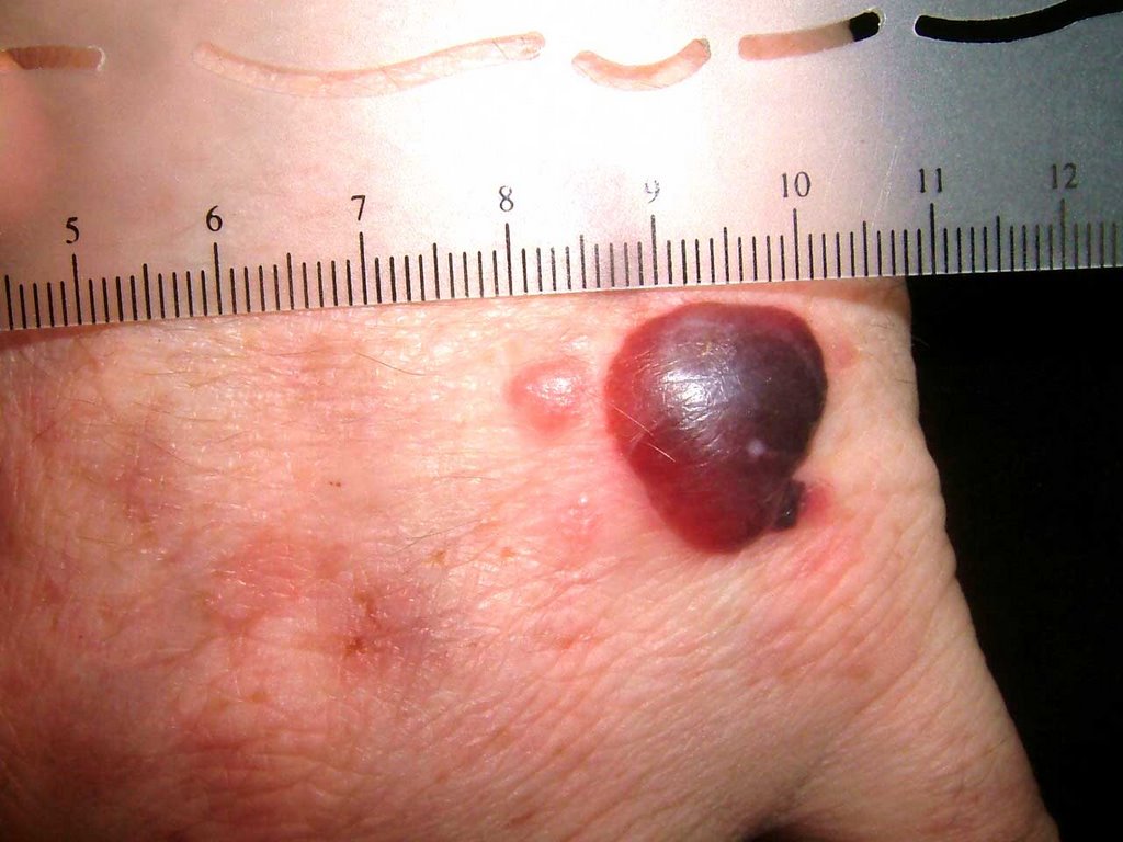
The patient is a 28 yo man referred on a walk-in basis with an eight month history of a bullous eruption on the dorsum of his left hand. He gets one or two painless lesions a month. No history was sent over with the patient and he is a poor historian. His medications include Welbutrin, Clozeril and Soma (carisprodol/aspirin)


Exam:

The only findings are on the dorsum of the left hand. Here, there is a hemorrhagic bulla, a vesicle and areas of mild erythema at sites of previous lesions.


Biopsy:
I suspect fixed drug or pseudoporphyria cutanea tarda. I biopsied the small papule and the edge of the bullae and made a follow-up appointment.
Path Report:
A subepidermal separation, individually necrotic keratinocytes , and a sparse superficial perivascular lymphocytic infiltrate with occasional neutrophils .
NOTE : (A and B). Amphophilic globular material is seen deposited in the dermis and around blood vessels in both specimens. This material is P.A.S. stain positive, and stains negative for amyloid and elastic tissue. The differential diagnosis could include porphyria cutanea tarda , although this is usually less inflammatory. A subepidermal autoimmune bullous disorder could also be considered. An additional biopsy for direct immunofluorescence may be of help.


Laboratory Studiess:
CBC normal, LFTs normal, ferritin normal. Hep B and C negative, All urinary porphyrins well within normal levels. Uroporphyrin: 9.6 ug (nl < 30.0) Coproporphyrin 40 ug (normal < 65/24 hr)

Discussion: This is likely pseudoporphyria cutanea tarda. A similar case has been reported. Your comments are welcomed.

Sunday, November 5, 2006

Mystery Rash in Runners

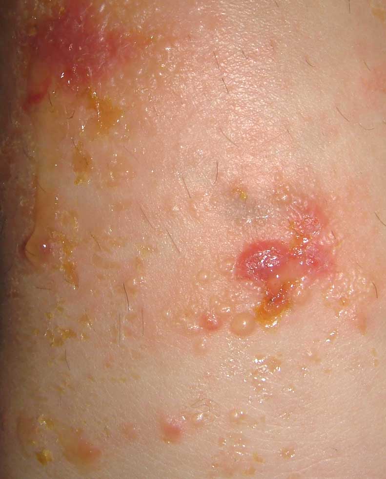
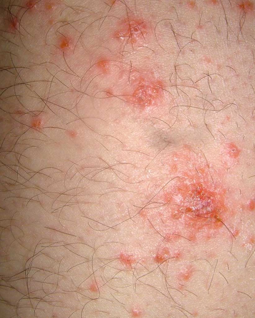
The Williams Cross Country running team went to a meet on the Connecticut shore on Saturday, October 28. There had been a big storm the previous day. The course took them through brackish water sometimes thigh deep. Within a 24 - 48 hours, around half of the runners had developed intensely pruritic lesions on arms and legs. The vast majority of the lesions were on exposed skin. The primary lesion appears to be a juicy papule or a clear vesicle. Some of these have crusted over in a few days. The lesions are discrete. They do not have the linear pattern characteristic of phytocontact dermatitis from a plant such as poison ivy. The pictures were taken of the legs of an 18 yo woman and a 21 yo man. The lesions were quite similar.


It's likely that this is from something dispersed in the brackish water they ran through. Runners from other schools were affected as well. About 50 runners from Williams College developed the eruption.

The possibilities include:
Atypical plant contact dermatitis (Some plant toxin such as urushiol churned up by the storm and floating in the water.)
Jellyfish nematocysts free floating after the storm and blown in
Bird schistosomes -- this is severe for that.
Bacterial infection (a culture was taken from one student and results were not specific)

Do you have other thoughts? What is your opinion?

Solution: The Cross Country course was redirected after the storm. A new path was cut though brush to circumvent a brackish area. It is likely that the new path went through poison ivy. The appearance of the rash 24 - 48 hours after exposure in the majority of runners and the quality of the itch suggests rhus contact dermatitis.

Sunday, October 22, 2006

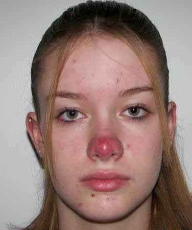
Teenager with a Red Nose


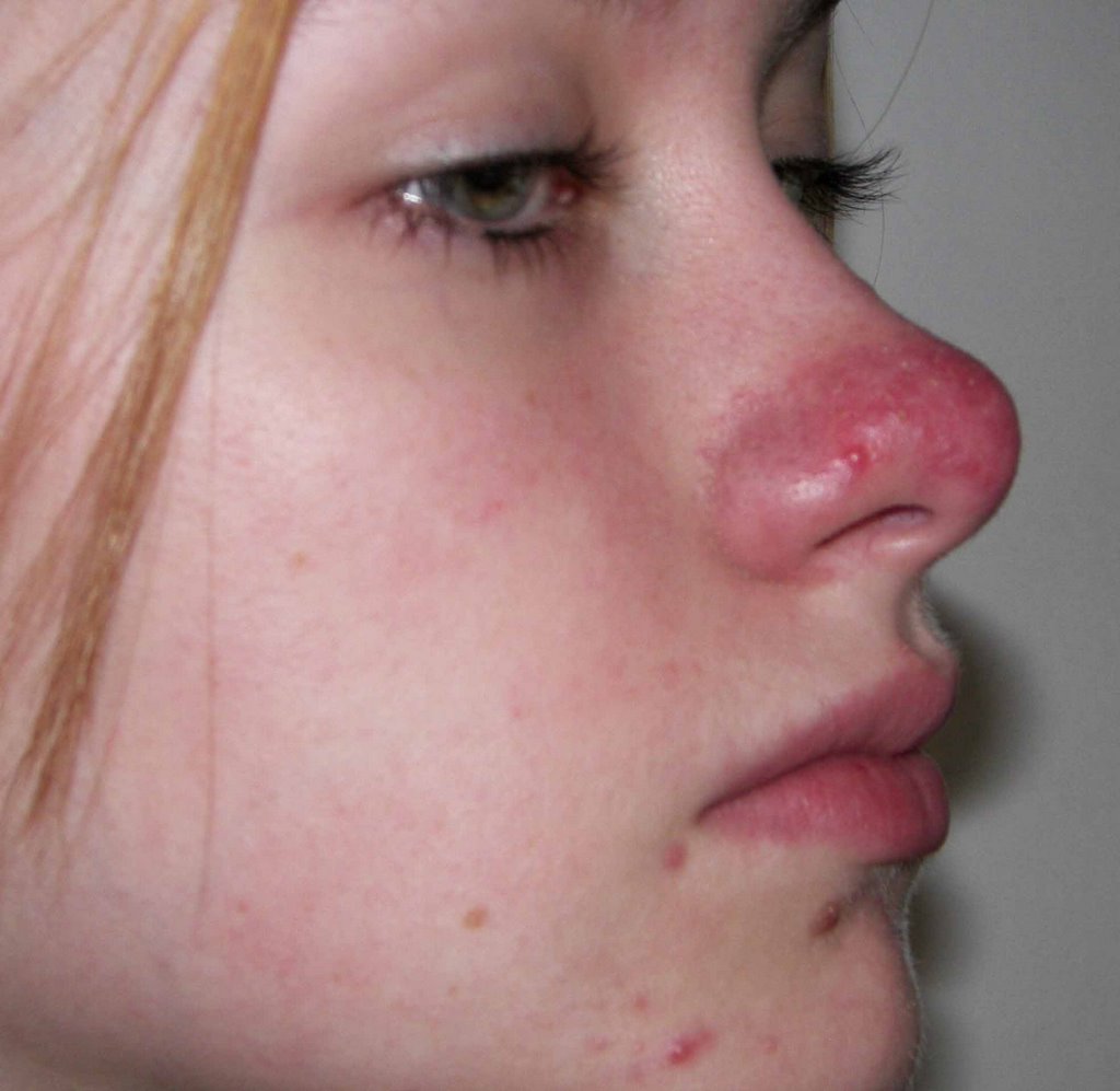
This 14 year-old girl has had a red nose for the past two years. It followed as severe sunburn. Her nose never looks normal. Occasionally, there are small cysts with bleeding and purulent drainage.


I have seen this picture occasionally in teenagers. It looks like a rosacea variant. I'd also consider demodex.

Your suggestions are anticipated and will be welcome.

Sunday, September 24, 2006

30 year old woman with changing mole


This 30 yo woman has noticed an enlarging pigmented lesion in her right axilla for almost a year. Her mother, a registered nurse, asked her to see a dermatologist. Her husband did not notice it.
The patient has Type II skin. The general cutaneous exam was unremarkable save for a 1.2 cm in diameter barely elevated plaque. There is a play of pigment and outline is irregular.

An excisional biopsy was performed.

Pathology: Malignant melanoma in situ , superficial spreading type. (Read by H. Byers of BU Skin Path who took photomicrographs) "The specimen exhibits a marked nested and lentiginous melanocytic proliferation with large severely atypical epithelioid cells. There is irregular nesting, focal confluent lentiginous melanocytic proliferation and cellular dyshesion."


She is scheduled for a wider excision, 0.5 cm on either side. No further work-up other than regular follow-up visits.

Your thoughts are appreciated.

Tuesday, September 19, 2006

Positive Band Aid Sign

Most dermatologists know about the "positive band aid sign." To me, it means a skin cancer has been lurking there for a while.

This 82 yo man came in with a one year history of a "sore" on his back. The drainage stained his shirt. When the band aid was removed, there was a one cm in diameter clean friable tumor, most likely a basal cell.

The lesion was shave biopsied and desicated and curetted. I'll affix a copy of the biopsy report when I get it.

How many of you use the term "Positive band Aid Sign?"