This is default featured slide 1 title

Go to Blogger edit html and find these sentences.Now replace these sentences with your own descriptions.

This is default featured slide 2 title

Go to Blogger edit html and find these sentences.Now replace these sentences with your own descriptions.

This is default featured slide 3 title

Go to Blogger edit html and find these sentences.Now replace these sentences with your own descriptions.

This is default featured slide 4 title

Go to Blogger edit html and find these sentences.Now replace these sentences with your own descriptions.

This is default featured slide 5 title

Go to Blogger edit html and find these sentences.Now replace these sentences with your own descriptions.

Wednesday, February 2, 2011

Unusual Nail Bed Tumor

Abstract: 67 yo man with 1 - 2 month hx of a nail bed tumor.

HPI:
67 yo man with 1 - 2 month hx of a nail bed tumor. The overlying nail has been destroyed. Lesion is asymptomatic.

O/E: 8 mm diameter flesh-colored tumor left thumb nail. No pigment. Lesion is solid, not friable.

Clinical Photos


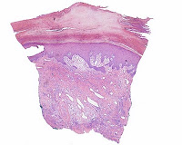
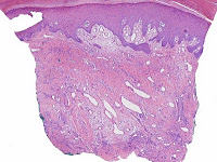
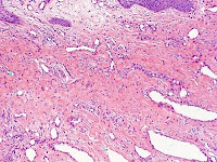
Pathology: Hyperkeratosis, parakeratosis, epidermal hyperplasis. There are ectatic blood vessels in an edematous and fibrotic stroma. Iin the DDx is an unusual traumatized hemangioma or subungual fibroma.

Microscopic Photos
courtesy of Deon Wolpowitz, Boston University SkinPath


Diagnosis: Benign nail bed tumor, not otherwise specified. Presented for ideas.

Questions: What is your diagnosis, and how would you approach this lesion?

Plan: The patient has been referred for defintive surgery to a Mohs surgeon.

Saturday, January 29, 2011

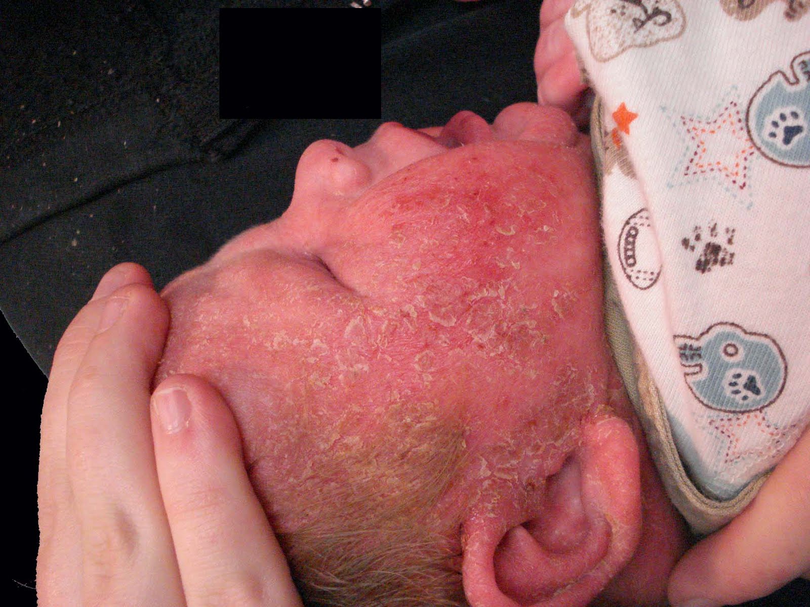
Dermatitis and Failure to Thrive

Abstract: 3.5 month old male infant with Failure to Thrive and Dermatitis

HPI: This 3.5 mo old infant male is the product of a normal pregnancy and delivery. He has had a severe dermatitis since shortly after birth. The process is most prevalent on head and neck and torso. He has lost weight on 150 kcal/kg per day. On visits seems happy and content. Three older siblings are all normal.

O/E: Widespread scaly patches on trunk and scalp. Thick cradle cap, greasy scale scalp and face. Background erythema. Red excoriated napkin area.

Photos: Taken when child was 9 weeks old.


Labs: Hi K+ and NA on hospital admission for FTT (but since normalized). Mild eosiniophilia and increased plts. Normal: CBC, ZN, IgE, IgM, IgA, IgG, amino acids. Normal karyotype, negative FISH for Williams Syndrome. Complement levels will be done and hair will be looked at for trichorhexis invagninata.

Diagnosis: Unclear. Initially I thought this child had infantile seborrheic dermatitis, but that usually responds well to treatment. At this point, Leiner's and Netherton's syndromes need to be ruled out. While neglect was initially considered, the child's pediatrician feels at this time that the mother is competent and has raised three other normal children.

Discussion: I saw this child once six weeks ago and because of transportation problems they could not keep f/u appointments. The eruption was originally treated with HC valerate 0.2% cream. Scalp hygiene was discussed. At this point a systemic process is considered.

Questions: In addition to considering Netherton's and complement deficiency syndromes like Leiner's what else would you recommend?

Follow-Up: The patient saw a pediatric gastroenterologist who changed her formula to one that excluded all milk proteins -- the eruption cleared completely in a few days and weight gain ensued. This was a milk allergy that masqueraded as a serious underlying disorder.

Sunday, January 9, 2011

Brachioradial Pruritus

We continue to get questions from readers -- but many do not leave an email address. If you want specific questions answered, I will try to do so but need an email to respond to. As of March 18, 2012, we've had 68 comments to this post -- and some people ask specific questions. If you want a response, you will have to include your email address. Otherwise, we have no mechanism to reply. We do not share email addresses with anyone. We do not bill anyone for anything.

June 15, 2012.  NEW LINK:  WE HAVE POSTED AN INFORMATION SHEET ON DermatologyCentral.  It's a work in process but should be helpful:  Brachioradial Pruritus Resource Page.
 
I have had an interest in Brachioradial Pruritus (BRP) for over 25 years and published a paper on it in 1985 and co-wrote a chapter on BRP for eMedicine.com. (Actually, the lion's share of the writing was done by Julianne Mann, then a dermatology resident at Oregon Health Sciences University.) As a result of the eMedicine.com chapter, I receive a few requests for information each year. Two came in recently and, with their permission, are presented here. Perhaps, some of our readers have suggestions for management of this annoying and occasionally disabling problem. Note: It is amazing how many comments have been made by people who suffer with BRP. Around 55 at this time. If you wish to leave a comment, and would like us to acknowledge it, please send me your name and email address. It will not appear anywhere on this site; but if you want me to relpy to you I need your contact information. Thank you, DJ Elpern.

1. This is from a 32 y.o. equestrian
I am an otherwise healthy woman desperately searching for a doctor who may be able to discuss some treatment options for my very itchy arms! I am 32 year old, 110 lb, active, female resident of New England and have been suffering with itchy arms (without rash) for at least four years now. When I finally sought treatment- I was referred by my primary MD to a dermatologist who informed me that I had dry, sensitive skin, and prescribed a topical cream and an oral steroid - I carefully followed his advice for 2 years - with absolutely no relief from the itch. The only thing that relieved the symptoms (which flare more intensely in the evening hours) was ice packs. I was often woken from sleep by the itch.

The itch occurs in cyclical pattern- symptomatic for months, followed by a month or two of none or very mild symptoms, then followed by another flare. it is present in sunny months as well as winter.

Finally, frustrated by the unresolved symptoms, at my own expense, I had one visit with a dermatologist in a nearby large city who very quickly diagnosed brachioradial pruritus and prescribed topical Capsaicin. Capsaicin works - sort of. But, it is a total pain in the butt for minimal relief. I am seeking alternative treatment.....acupuncture, chiropractic, anything ... ? Do I need an X-ray? What do I do? I am on my own out here in a sea of medical professionals who seem unwilling to take this condition seriously or look for possible causes that may be an underlying cause for this condition of itchy arms....

It may also be of interest that I have a large amount of muscle/ligament type tension in my neck and shoulders- it's pretty severe, some of this tension is a manifestation of stress, some results from my very physical occupation as an Equestrian.... I am personally tempted to think that all the tension in my neck and shoulders may have something to do with the arm itch - but this conflicts with the cyclical nature of the itch. Has the tension is chronic?
To me, BRP it's a big deal!


2. This is from a 37 yo health care professional:
I have suffered with this for no less than 5 years. Until about a year ago it was sporadic, but has been substantially worse and constant for the last year or so. Nights are particularly difficult, as the itching becomes intolerable and uncontrollable. I have recently found some comfort with ice packs, but naturally having to traipse off to the kitchen several times a night is inconvenient and exhausting. The most recent prescriptions I have tried are 10mg cetirizide for daytime and 25mg doxepin (1-2 at bedtime), and also betamethosone cream. The cetirizide helps with other allergy issues I have, but doesn't offer any relief to the itchy arms. Also, the doxepin helps me get a few solid hours of sleep, if I take 2, but, as with hydroxizine, I feel "hungover" the next morning... AND insatiably hungry.
I do LOVE the sun, and have always found that I feel better through the cold months if I go tanning a couple times a week. In the winter months, I spend a bit of time tanning prior to spending any significant time outside in the summer. I have fair skin, and it helps prevent burning before spending a day at the beach. (which I do, ALL day, as often as possible.) I do not, however notice any increase or decrease in the itching based on exposure to sun/tanning. In the last year, however, I HAVE starting being a bit more careful and use a minimum of SPF 30 during peak hours at the beach. Here is a recent picture:
(Editor's note: This shows some typical findings. The skin looks a bit dry and lichenified and there is evidence of excoriation. Not all cases show these changes, some are more subtle.)


I have seen several doctors, including a dermatologist, regarding this debilitating itch and NOTHING has helped enough to warrant the side effects. I cannot seem to get anyone to understand that it isn't like a skin itch, it feels like its UNDER my skin. I scratch so much, especially at night, that I bleed. I am not a crazy, irrational person.... but the loss of sleep and inability to find anything that relieves this itching probably makes me seem as though I am.

Comment: Both of these patients appear to have chronic BRP. I see a similar patient once or twice a year. While most patients with BRP have episodic, relatively easy to treat disease, there are a few patients who have disabling symptoms. There is an aphorism, "It is often more important to treat the patient who has the disease than the disease the patient has." I think this applies to persons with chronic BRP. In a real way, these are orphan patients. Few dermatologists have the expertise, and fewer the time, to adequately evaluate and treat these patient. If you have suggestions, they would be most appreciated.

Actual Use
Most patients with BRP will get some relief from ice packs.  3M makes a reusable pack thatsells in the U.S. for $2.50.  This is an inexpensive way to start treating BRP if you have not used ice already.  You could use this 2 - 3 times a day for 15 - 20 minutes or whatever works best for you.

Friday, December 31, 2010

ULE?

Abstract: 4 yo girl with 3 week hx of papular eruption left axilla
HPI: This otherwise healthy four year old girl has had a mildly pruritic papular eruption which began in the left axilla with a dermatitic appearance and spread centrifugally where it appeared more papular. There was no antecedent illness. The eruption was first seen on 12/23/2010, cleared after a few days and they reoccurred. The child is not bothered by it. No animals at home and no other family members similarly affected.
O/E: Discrete erythematous papules in the left axilla and surrounding areas. The individual lesions are 3 - 5 mm in diameter. The right axilla and thoracic area are clear. There is left axcillary adenopathy
Clinical Photos: 12/31/2010
Left Axilla

Right Axilla
Lab: none

Diagnosis:
Unilateral Laterothoracic Exanthem.

DDx: The lesions look like bites. Against that is the lack of symptoms, the unilateral location, and no likely cause of bites. Contact dermatitis seems unlikely.

Discussion: This is an unusual entity but the child seems well otherwise and I have recommended no treatment for present. I have not seen ULE before (to my knowledge); yet this seems the likely diagnosis. Unfortunately, the pictures in the textbooks are not very good. I suspect, that like Gianotti-Crosti, ULE may be caused by a number of viruses and that it will be difficult to come up with an etiology. The clinical appearance seems to be protean, but the distribution aids in making a diagnosis.

Reference:
1. Emedicine.com has a good chapter, however, they use the name "Asymmetric Periflexural Exanthem of Childhood."

2. McCuaig CC, et. al. Unilateral laterothoracic exanthem. A clinicopathologic study of forty-eight patients. J Am Acad Dermatol. 1996 Jun;34(6):979-84.
Department of Pediatrics, Hôpital Sainte-Justine, Montreal, Quebec, Canada.
Abstract
BACKGROUND: Four years ago, we began seeing young children with an unusual, predominantly unilateral, morbilliform and eczematous, self-limited cutaneous eruption. It appeared to correspond to unilateral laterothoracic exanthem (ULE) reported from France and to an eruption described as "a new papular erythema of childhood" in the United States.
OBJECTIVE: We conducted a prospective study of ULE to define its clinical evolution, pathology, and therapy. In addition, we performed epidemiologic and microbiologic investigations in an attempt to determine the cause of ULE.
METHOD: We studied 48 children with ULE. In some patients, blood, urine, stool, as well as skin biopsy specimens were analyzed.
RESULTS: ULE is a morbilliform, eczematous eruption that often begins close to the axilla and spreads to become bilateral, although it usually retains a unilateral predominance. Patients' mean age at onset is 24.3 months, with a female predominance (2:1) and mean duration of 5 weeks, followed by spontaneous resolution that may or may not be improved with topical corticosteroids. It is characterized by a unique eccrine lymphocytic infiltration. Although signs of infection were reported by most patients, no one infectious agent was identified. No significant epidemiologic factor was found.
CONCLUSION: ULE, in young children, is a self-limited morbilliform and scarlatiniform eruption that may represent a specific skin reaction to one or more infectious agents.

Follow-up Note: The patient's symptoms continued to wax and wane and she was seen by a pediatric dermatologist. At that time, there was just one lesion in the left axilla ( see picture) and a diagnosis of psoriasis was made. There were no other stigmata for psoriasis. If this is the case, the initial picture did not suggest that. Since the sole lesion present now is a plaque in the left axilla, if this turns out difficult to control with topical steroids, consideration to using tacrolimus should be given.


References
:
Tacrolimus ointment is effective for psoriasis on the face and intertriginous areas in pediatric patients.
Brune A, Miller DW, Lin P, Cotrim-Russi D, Paller AS. Pediatr Dermatol. 2007 Jan-Feb;24(1):76-80.
Abstract
Children with psoriasis often have involvement of the face and intertriginous areas. While corticosteroids have been the mainstay of treatment for plaque-type psoriasis, the face and intertriginous areas are more sensitive to local effects of topical steroid use such as cutaneous atrophy. Topical tacrolimus has shown promise in adult patients as an alternative antiinflammatory without the cutaneous side effects of steroids. Eleven patients between 6 and 15 years of age with facial or inverse psoriasis were evaluated in a 6-month, single-center, open-label trial. Clinical evaluations were made at baseline and days 30, 90, and 180. Severity was assessed using the physician's global assessment of improvement relative to baseline, a 6-point rating scale for signs of disease (erythema, infiltration, desquamation), and an overall severity score. Within the first 30 days of treatment, every patient had cleared or achieved excellent improvement with the use of tacrolimus ointment. Statistically significant improvement was achieved in each sign of disease and the overall severity score. The only adverse event reported in 6 months of observation was significant pruritus in one patient. We therefore conclude that tacrolimus ointment is an effective treatment for psoriasis on the face or intertriginous areas in children.

Wednesday, December 22, 2010

Dermatomal Eruption

Abstract: 39 yo man with two month history of dermatomal eruption.
HPI: This 39 yo man developed a dermatomal vesicular eruption 2 months ago. He was seen by his GP and treated with valcyclovir and it cleared somewhat but not completely. The eruption continues to evolve. He complains of pain and pruritus. Feels well otherwise. No underlying diseases known of.
O/E: There is a dermatomal process extending from T-10 to L 2 on the right side. The lesions are scaly patches. There are no vesicles. The lesions do not cross the mid-line. Area biopsied today is only a couple of days old, by history.
Photos:


Diagnosis:
Atypical Herpes Zoster. H.z. progressing over a two month period (especially after valcyclovir) is quite unusual in a healthy, immunocompetent person.

Plan: I did a biopsy and checked his chemistries and CBC. I have seen patients with HIV/AIDS and a patient with angioimmunoblastic lymphadenopathy with atypical HSV and HZ; but never a patient like this. Perhaps, this is something I am not thinking about. I will post a photomic when path is reported and lab results. For the time being, I prescribed acyclovir 800 mg 5 times a day.

Addendum: Fran Storrs felt this was an eczematous process, possibly a contact dermatitis. The pathology showed no multinucleated giant cells and had features of a "dermatitis." So, was this a dermatitis secondary to H.Z., an atypical contact dermatitis, or factitial (the patient did ask for pain meds when first seen, which were not given)? He is now being treated with a topical corticosteroid now and we'll see how he does. When seen for suture removal sight days after biopsy, the eruption looked a bit better, was less symptomatic and had not spread beyond the dermatomes first involved.


Sunday, October 17, 2010

The Power of BLINCK

Presented by Yoon Cohen MS IV, University of New England, Biddeford, Maine and David Elpern MD, Williamstown, Massachusetts

Abstract: 68 yo woman with 4-6 months history of an atypical melanocytic lesion.

HPI: This healthy 68 yo woman with type II skin presented to the clinic with 4-6 months history of an atypical melanocytic lesion on the left knee. She had noticed an increase in size and change in color and was concerned about these changes in the lesion.

O/E: There was a 7 mm in a diameter asymmetrical brownish macule on the left knee. The lesion showed an irregular border with varied colors.

Clinical photographs:


Dermoscopic images:


Microscopic images:
Dermatopathology report:
The specimen exhibits a proliferation of moderate to severely atypical melanocytes distribubted in irregular nests, as well as singly at and above the dermal epidermal junction, with pagetoid spread to the granular layer and near confluence over at least three rete ridges. These findings support the histologic diagnosis of melanoma-in-situ.

Our appreciation to Dr. Deon Wolpowitz, MD from Boston Univeresity, Dermatopathology, for providing these photomicrographs for the case.

4X


10X


20X


20X


Diagnosis:
Malignant melanoma in situ

Discussion:

The BLINCK approach:

We followed the BLINCK checklist, introduced by Dr. Peter Bourne, a founder of the Skin Cancer College of Australia and New Zealand (SCCANZ). The score for the lesion was added up to 4 by criteria as following.

1. B. The lesion was not clearly benign at our first initial evaluation
2. L. The lesion appeared to be lonely without any other similar melanocytic lesion near by
3. I. The lesion appeared to be irregular outline and color on our dermoscopic exam
4. N & C. The patient was nervous about the change in color in past 4-6 months
5. K. The lesion exhibited known clues when viewed with a dermatoscope. See "Chaos and Clues" reference below.
BLINCK Score = 4

According to the BLINCK approach, a lesion should be biopsied if the BLINCK score is 2 or more out of a possible 4. Therefore, the we excised this lesion and sent for a pathologic evaluation.

According to Dr. Bourne, the BLINCK approach is presented as a simple method to assist the clinician with the decision of whether to biopsy a skin lesion or not. The use of this algorithm will improve the pickup rate of potentially serious skin cancers as well as reduce the number of unnecessary benign lesion excisions. BLINCK may be especially helpful to clinicians who have only basic or intermediate dermoscopy skills but who are regularly called upon to assess skin lesions in their practices.

Questions:
1. Would you consider using the BLINCK approach at your practice?
2. If you already have adapted the BLINCK approach, how have your experiences been?

References:
1. McColl I. BLINCK. http://idsblinck.blogspot.com/2009/11/blinck.html. Updated November 19, 2009. Accessed September 7, 2010
2. Rosendahl C, Kittler H, Cameron A, et al. CHAOS & CLUES - The Algorithm. http://www.chaosandclues.blogspot.com. Updated November 19, 2009. Accessed September 7, 2010

Tuesday, September 21, 2010

Palmoplantar Erythrodysesthesia Syndrome

Presented by Yoon Cohen, MS IV
University of New England College of Osteopathic Medicine


Abstract: 45 yo woman with a metastatic breast cancer and a painful hand/foot dermatitis.

HPI: This is a 45 yo woman who was diagnosed with a metastatic breast cancer a few years ago. She has developed "atopic dermatitis" like symptoms on her hands and feet since starting capecitabine. She has completed 5 cycles of Xeloda (capecitabine) and has had to reduce the dose because of this condition. The lesions started as dry edematous, erythematous areas on palms and soles with a tingling sensation.

O/E: The examination reveals general areas of desquamation and hyperlinearity with mild erythema on both palms. This is best shown on the first photograph. 

Clinical photographs:


Diagnosis: Palmoplantar erythrodysesthesia syndrome, hand-foot syndrome, chemotherapy-induced acral erythema

Discussion:
Chemotherapy-induced acral erythema or palmoplantar erythrodysesthesia syndrome is a well-defined reaction to some of the chemotherapeutic agents such as methotrexate, cytarabine, doxorubicin, fluorouracil, cytosine arabinoside, and bleomycin. This reaction is characterized by symmetric, well-demarcated, painful erythema of the palms and soles, which may progress to desquamation or blisters. It appears to be dose dependent, and is likely a direct toxic effect of the drug. Tingling on the palms and soles is followed in a few days by painful, symmetric, well-defined swelling and erythema [4].

Questions:
1. Please feel free to share your experiences with treatment options.

References:
1. Marini A, Hengge UR. Hand-foot syndrome with capecitabine therapy. Hautarzt. 2007 June; 58(6):532-6
Abstract: A 72-year-old patient with esophageal carcinoma developed a severe hand-foot syndrome during second-line therapy with the oral fluoropyrimidine capecitabine. We also summarize the current knowledge with regard to the hand-foot syndrome and distinguish it from palmoplantar erythrodysesthesia.

2. Degen A, Alter M, Satzger I, et al. The hand-foot-syndrome associated with medical tumor therapy- classification and management. J Dtsch Dermatol Ges. 2010 Sep; 8(9):652-61
Abstract: The hand-foot-syndrome (HFS, palmoplantar erythrodysesthesia, chemotherapy-associated acral erythema) is characterized by painful predominantly palmo-plantar lesions. The association with different chemotherapeutic agents has been known for over 20 years. More recently, HFS has been reported in association with regimens using targeted agents, in particular the multikinase inhibitors (MKI) sorafenib and sunitinib. The HFS associated with MKI has a different distribution and clinical appearance than the traditional disorder. In this review, similarities and differences between chemotherapy- and MKI-associated HFS are discussed and current recommendations for their prophylaxis and management are summarized.

3. Janusch M, Fischer M, Marsch WCh, et al. The hand-foot syndrome - a frequent secondary manifestation in antineoplastic chemotherapy. Eur J Dermatol. 2006 Sep-Oct; 16(5): 494-9
Abstract: The hand-foot syndrome (HFS) (palmoplantar erythrodysesthesia) designates acute, painful erythemas of the palms and soles of the feet caused by antineoplastic chemotherapies. The most frequent trigger substances are 5-fluoruracil and its derivates. At maximum severity, the HFS is bullous to erosive or ulcerous in character. The pathogenesis has not yet been clarified. Histologically, the HFS is characterized by a toxic keratinocyte reaction. Furthermore, there is sub-basal edema with a tendency to bullae, dilated blood and lymph capillaries and usually only mild perivascular lymphocytic infiltration. Early recognition and delineation from other differential diagnoses is prerequisite to targeted management of the disease. Depending on the severity, HFS requires dose reduction, interruption or switch in the antineoplastic chemotherapy. (You can access to the article at http://www.john-libbey-eurotext.fr/e-docs/00/04/26/D7/vers_alt/VersionPDF.pdf)

4. Habif TP. Clinical Dermatology. 5th edition. USA: Elsevier Science 2010