This is default featured slide 1 title

Go to Blogger edit html and find these sentences.Now replace these sentences with your own descriptions.

This is default featured slide 2 title

Go to Blogger edit html and find these sentences.Now replace these sentences with your own descriptions.

This is default featured slide 3 title

Go to Blogger edit html and find these sentences.Now replace these sentences with your own descriptions.

This is default featured slide 4 title

Go to Blogger edit html and find these sentences.Now replace these sentences with your own descriptions.

This is default featured slide 5 title

Go to Blogger edit html and find these sentences.Now replace these sentences with your own descriptions.

Saturday, August 10, 2013

Hemorrhagic Blister


Abstract: A 58-year-old man with 2-day history of deep purple to black papule

History: A 58-year-old man presents for evaluation of a skin lesion on the right lower abdomen that has been present approximately 2 days. The patient does not remember having any trauma on that area. It seems the lesion appeared quite abruptly and the color has been darkened over past 2 days. No history of non-melanoma skin cancers or melanoma.

O/E: The skin exam shows a healthy and pleasant man with skin type III. There is a sharply demarcated 6 mm deep dark purple oval shaped papule on the right lower abdomen. It is somewhat compressible.

Clinical & Dermatoscopic Photos:

Polarized Dermatoscopic Photo
Non-Polarized Dermatoscopic Photo

Diagnosis: Hemorrhagic blister

Discussion: The lesion was lanced with an 11 blade. Blood product was drained and confirmed the diagnosis of hemorrhagic blister. Initially, the lesion did not obviously look as  a hemorrhagic blister. The dermatoscopic image did not show a typically appearing hemangioma. Our biggest concern was to make sure this is not a melanoma. Conventionally, melanoma would not appear in a 2-day period. A careful history led us to consider a hemorrahagic blister with a simple lancing. 

Reference:
1. Bullous malignant melanoma: an unusual differential diagnosis of a hemorrhagic friction blister.
Vogt T, Brunnberg S, Hohenleutner U, et al. Dermatol Surg. 2003 29(1): 102-4

Source

BACKGROUND:

A 66-year-old woman presented to our outpatient clinic with a 3 x 2.5-cm tense, hemorrhagic-appearing bulla on her forefoot. Histopathology and immunohistochemistry confirmed a transtumoral-transepidermal blister formation within an advanced acrolentiginous malignantmelanoma (MM).

OBJECTIVE:

To study bullous malignant melanoma.

METHODS:

Blistering in MMs represents a rare but clinically important pitfall in clinical differential diagnosis. The blisters are typically due to the disruption of the cohesion between neoplastic cells and keratinocytes, but physical friction may also contribute.

RESULTS:

Hemorrhagic blistering is, in many cases, a relatively insignificant finding in which frictional forces are imposed.

CONCLUSION:

The case reported here underscores that in rare cases MMs, particularly if acrally located, can be complicated by hemorrhagic blistering. Because of the life-threatening consequences, one should be aware of this rare differential diagnosis.


Sunday, August 4, 2013

Rosacea-Like Demodeciasis

Abstract:  47 yo woman with ab 8 month history of a facial eruption
 
The patient is a 47 yo woman with a rosacea-like eruption for eight months.  Dr. Yoon Cohen suggested demodicosis and performed a scraping.  It was loaded with demodex mites.  We are considering this an example of rosacea-like demodexinfestation and treated the patient with ivermectin 250 microgram/kg x 2 a week apart and permethrin 5% cream 2 - 3 times a week. 

Photos:

 After one week:

Diagnosis:  Rosacea-like Demodeciasis (most probably)

Discussion: It seems that this acneiform eruption is due to Demodex, but we need more time to be certain. Since Samuel Ayers, Jr. described this entity in 1930 it has been in the literature but there are still doubters in the dermatologic community.

Reference:


1. Dermatol Online J. 2007 Oct 13;13(4):9.
Granulomatous rosacea-like demodicidosis. Free Full Text
Lee JY, Hsu CK.
Abstract
Demodicidosis may present as pityriasis folliculitis, papulopustular lesions, rosacea-like eruptions, and granulomatous rosacea-like eruptions. We report a case of demodex granuloma presenting with recurrent granulomatous rosacea-like papules on the face in a middle-aged woman. The diagnosis of demodicidosis was made by finding extrafollicular mites in the perifollicular inflammatory infiltrate. The papules resolved after 3 weeks of systemic and topical metronidazole, and low-dose oral prednisolone therapy. In summary, demodex granuloma may be mistaken for granulomatous rosacea-like papules. Correct diagnosis can be facilitated by finding extrafollicular demodex mites in skin biopsy specimens.

2.  Cutis. 2007 Aug;80(2):149-51.
Recalcitrant papulopustular rosacea in an immunocompetent patient responding to combination therapy with oral ivermectin and topical permethrin.  Full Text PDF
Allen KJ, Davis CL, Billings SD, Mousdicas N.
Abstract
A 68-year-old healthy man presented with papulopustular rosacea (PPR) recalcitrant to multiple therapies, including permethrin cream 5%. Histologic examination detected the presence of chronic folliculitis and numerous Demodex organisms. A diagnosis of rosacealike demodicidosis was rendered, and the patient was treated with oral ivermectin and permethrin cream 5%, resulting in resolution of the folliculitis. Demodex infestation should be considered in any patient with rosacealike dermatitis resistant to conventional rosacea therapies. If infestation is demonstrated in these patients, oral ivermectin in combination with topical permethrin is a safe and effective therapeutic option.

Follicular Mucinosis?


Abstract: 80 yo man with intensely pruritic papules face and neck for 2 months

HPI:  This 80 yo man has a 2 month history of 2 – 3 mm erythematous papules, mostly on face and neck. These are few in number.  A trial cobetasol ointment was without relief.  His medications are: amlodipine, HCTZ, metroprolol, Vytorin (ezetimibe and eimvastatin), Pantoprazole.  He is in good general health.

ClinicalPhotos: These show how subtle the lesions are.


Because of symptoms a Biopsy was done.
Histopath shows: focal erosion and ulceration and ectatic blood vessels.  There is a moderate to dense superficial and deep perivascular perifollicular lymphohistiocytic infiltrate with numerous eosinophils, marked mucin deposition and conspicuous disruption of the follicular epitheliumThe pathologic diagnosis includes, “infectious etiology,” eosinophilic folliculitis, and follicular mucinosis.

Diagnosis:  Acneiform Follicular Mucinosis might be the best fit.

Reference:


Sunday, July 21, 2013

Chronic Ulcers

Presented by Yoon Cohen, D.O.
Alta Dermatology Residency
Mesa Arizona

Abstract: 66-year-old woman with 3 year history of leg ulcers

HPI: The patient is a 66-year-old retired elementary school teacher who was seen for evaluation of chronic leg ulcers. The most recent injury was sustained on her right lower leg when she bumped into her grandson’s toy.  This  eventually caused a new leg ulcer. Subsequently, she was hospitalized, and seen and evaluated by the Plastics Wound Clinic at a major hospital center. The patient presented for continuity care after discharge from the hospital.  Her chronic leg ulcers started in 2011 when she had a cat scratch on the right foot which resulted in cellulitis and ulcer on the right leg. After that episode, she noticed the development of ulcers whenever she gets even minor injury to her legs. She has a long history of rheumatoid arthritis that she has been on multiple treatments including prednisone, methotrexate and Remicade. Her current medications include prednisone 5mg daily, methotrexate 20mg weekly with folic acid, Celebrex 200mg bid, and oxycontin 50mg daily.

O/E: The skin exam shows a pleasant, somewhat depressed woman with several various seized leg ulcers with healthy granulation tissues at base on the lower legs.
A) R lateral calf: fibrinous base, 3.3 cm x 1.3 cm
B) L dorsal foot: Fibrinous base, 1.8 cm x 1.0 cm
C) L lateral calf: Fibrinous base, 2.5 cm x 1.6 cm
D) L medial calf: Clean granulation tissue at base, 5.0 cm x 4.8 cm x 0.5 cm
Pedal pulses 1+ and 2+ pitting edema bilaterally

Clinical Photos:


Hospital Courses:
1. Diagnosis: Her chronic ulcers were considered to be multifactorial. Likely contributors include trauma, infection, rheumatoid arthritis, venous insufficiency, ulceration from methotrexate toxicity.
2. Procedures: Debridement and Kenalog injection(10mg/cc) to the left medial calf
3. Diagnostic Studies: Wound culture (pending)
4. Dressings: Acticoat 7 for lesions A and D, and then continue hydrofera blue to lesions A and D. Santyl, adaptic, gauze, kerlex for lesions B and C.
5. Compression: Soft cotton between the skin and the SurePress; will try penumoboots for home and leg elevation.

DiscussionThe possible etiologies can be trauma, rheumatoid arthritis, venous insufficiency, infection, or prolonged use of methotrexate or Remicade. There are 9 reports of ulcerations from methotrexate and 1 report of ulcerations  of foot from Remicade (infliximab) according to Litt’s DERM Drug Eruptions & Reactions Manual. There are two major questions. What is (are) causing these ulcerations which occur with even minor injuries? Could it be methotrexate toxicity? Could this be a variant form of pyoderma gangrenosum related to RA? She does not heal well even in other locations of her body. It seems to be more than just venous insufficiency. Lastly, how can we help heal these ulcers? 

Questions: 
What are your thoughts regarding the etiology of this woman's leg ulcers? 
How would you manage her to facilitate healing?
Do you think her medications play a role?  or her rheumatoid arthritis?


References:
1. Ben-Amitai D, Hodak E, David M. Cutaneous ulceration: an unusual sign of methotrexate toxicity - first report in a patient without psoriasis. Ann Pharmacother. 1998; 32 (6): 651-3
Abstract
Objective: To describe a case of skin ulcer related to methotrexate toxicity
Case Summary: A 67-year-old white man with seronegative arthralgia developed recurrent skin ulcer concurrent with 3-year treatment with methotrexate 5 mg daily. The skin ulcer resolved on discontinuation of methotrexate
Discussion: To the best of our knowledge, this is the first reported case of skin ulcer believed to be induced by methotrexate in a patient without psoriasis
Conclusions: Skin ulceration should be added to the list of possible toxic adverse effects of methotrexate, not only in psoriatic patients but also in those without psoriasis.

2. Stolman LP, Rosenthal D, Yaworsky R, et al. Pyoderma Gangrenosum and rheumatoid arthritis. Arch Dermatol. 1975; 111(8): 1020-3. 

Abstract
Two patients had pyoderma gangrenosum and rheumatoid arthritis. Biopsy specimens of the cutaneous ulcers in the paitents showed a necrotizing vasculitis. Complement (C3) and immunoglobulines were not detected in the skin lesions. There was no important impairment of cell-mediated immunity observed.

3. Seitz CS, Berens N, Brocker EB, et al. Leg ulceration in rheumatoid arthritis -- an underreported multicausal complication with considerable morbidity: analysis of thrity-six patients and review of the literature. Dermatology. 2010; 220 (3): 268-73
CONCLUSIONS:
The majority of leg ulcers in patients with RA are due to underlying venous/arterial malfunction while vasculitic or traumatic ulcers are less common. Additionally, we identified a relevant subgroup of patients with 'inactivity ulcers' due to impaired mobility and consecutive lymphedema. Morphology and localization of ulcerations as well as duplex sonography provide the most important clues for accurate diagnosis, ensuring adequate treatment.

-->


Sunday, July 14, 2013

A potentially lethal disease

presented by Dr Henry Foong
Ipoh, Malaysia

Abstract:  19 yo man with skin lesions and gastro-intestinal infarcts

History: A 19 yr old student presented with multiple discrete mildly pruritic papules on the trunk and thighs of 9 months duration.  It started as papules on the trunk that become enlarged resulting a central depression.  2 months ago, he had severe abdominal pain,  underwent emergency laparotomy and was told he had a "perforated appendix".  The surgeon noted several shallow ulcers on the large and small intestines.  He was worried about typhoid but serology was negative.  

Exam:  Multiple discrete papules/plaques 3-6 mm diameter with a central porcelain white atrophied depression and a raised border on the trunk and thighs.  There was a rim of telangiectasia on the periphery in some of the lesions. No purpura was noted.

Clinical Photos:


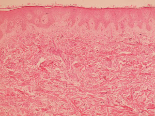
Pathology: A skin biopsy was performed.  Biopsy showed an atrophied epidermis with evidence of arteritis in the deeper dermis.  (see below)

Lab: His blood counts and biochemistry was unremarkable.

Diagnosis:  Degos disease (Malignant atrophic papulosis) appears most likely.  Could this some form of cutaneous lymphoma?

Questions:  Have any of our readers seen and treated similar patients? There are only around 200 cases of Degos Disease reported in the literature.  Treatment is difficult and often ineffective.  Aspirin and dipyridamole has been tried to reduce the complications of vessel thrombosis.  Some cases has been associated with connective tissue disease but by and large those with gastrointestinal complications have poor prognosis. IV immunoglobulins and eculizumab has been tried with some success.

Cost Considerations:  
In the U.S.: 

Eculizumab is ~$7000 per dose at low end

A four dose course of IVIG for a 70 kg person at 2 g/kg would cost $25 000-$26 000. Or ~ $21,000 for a 60 kg person. 

While these drugs may be 40% cheaper in Malaysia can your patient's family afford a trial or does the government pick up the cost?

References:
1. Cynthia M. Magro, MD, Jonathan C. Poe, PhD,et. al.  A C5b-9/Interferon-α–Mediated Endotheliopathy Syndrome.  Am J Clin Pathol 2011;135:599-610

2. Scheinfeld N. Commentary on 'Degos disease: a C5b-9/interferon-α-mediated endotheliopathy syndrome' by Magro et al: a reconsideration of Degos disease as hematologic or endothelial genetic disease.  Dermatol Online J. 2011 Aug 15;17(8):6. Excellent discussion and available Free Full Text 

3. Zhu KJ, et al. The use of intravenous immunoglobulin in cutaneous and recurrent perforating intestinal Degos disease (malignant atrophic papulosis) Br J Dermatol. 2007 Jul;157(1):206-7.

Histopath demonstrates an atrophied epidermis with evidence of arteritis in the deeper dermis.


Wednesday, July 3, 2013

Innoculation Eschar

Abstract: 6 yo  boy with one month history of an eschar on the neck

History:  This six year old boy has had an indurated nodule on the left posterior nuchal area for around a month.  He is a high functioning patient on the autism spectrum.  No history of antecedent trauma or envenomation is available.  He is well otherwise with no complaints of constitutional symptoms or headache.

O/E:  1 cm escharotic lesion left posterior neck.  Surrounding area is indurated.  No other cutaneous findings.

Clinical Photo:
photo courtesy of Yoon Cohen, D.O.
Labs:  A wound culture was obtained.

The eschar was sharply dissected.  We attempted to anesthetize the area for an incisional biopsy but patient was not compliant.

Discussion:  The patient lives in an endemic area for Lyme disease.  There are many ticks in the environment.  The wound may represent a reaction to a tick bite.  The differential diagnosis in patients with similar eschars includes rickettsial infections, cutaneous anthrax, tularaemia, necrotic arachnidism (brown recluse spider bite), scrub typhus (Orientia tsutsugamushi), rat bite fever (Spirillum minus), staphylococcal or streptococcal ecthyma, and Lyme disease.

Diagnosis:  Considering where this patient lives, the most likely diagnosis is tick bite reaction.

References:
1.  Tick-borne lymphadenopathy (TIBOLA) acquired in Southwestern Germany Free Full Text
Siegbert Rieg1 et. al  siegbert.rieg@uniklinik-freiburg.de
BMC Infectious Diseases 2011, 11:167 doi:10.1186/1471-2334-11-167

2.  African tick-bite fever: a new entity in the differential diagnosis of multiple eschars in travelers. Description of five cases imported from South Africa to Switzerland.
Althaus F, Greub G, Raoult D, Genton B.  Int J Infect Dis. 2010 Sep;14 Suppl 3:e274-6. doi: 10.1016/j.ijid.2009.11.021. Epub 2010 Mar 15.
Abstract:  African tick-bite fever (ATBF) is a newly described spotted fever rickettsiosis that frequently presents with multiple eschars in travelers returning from sub-Saharan Africa and, to a lesser extent, from the West Indies. It is caused by the bite of an infected Amblyomma tick, whose hunting habits explain the typical presence of multiple inoculation skin lesions and the occurrence of clustered cases. The etiological agent of ATBF is Rickettsia africae, an emerging tick-borne pathogenic bacterium. We describe herein a cluster of five cases of ATBF occurring in Swiss travelers returning from South Africa. The co-incidental infections in these five patients and the presence of multiple inoculation eschars, two features pathognomonic of this rickettsial disease, suggested the diagnosis of ATBF. Indeed, the presence of at least one inoculation eschar is observed in 53-100% of cases and multiple eschars in 21-54%. Two patients presented regional lymphadenitis and one a mild local lymphangitis. Though a cutaneous rash is described in 15-46% of cases, no rash was observed in our series. ATBF was confirmed by serology. Thus, ATBF has recently emerged as one of the most important causes of flu-like illness in travelers returning from Southern Africa. The presence of one or multiple eschars of inoculation is an important clinical clue to the diagnosis. It can be confirmed by serology or by PCR of a biopsy of the eschar. Culture can also be done in reference laboratories. Dermatologists and primary care physicians should know this clinical entity, since an inexpensive and efficient treatment is available.

3.  Histologic features and immunodetection of African tick-bite fever eschar. Free Full Text
Lepidi H, Fournier PE, Raoult D.    Emerg Infect Dis. 2006 Sep;12(9):1332-7.
African tick-bite fever (ATBF) is a rickettsiosis caused by Rickettsia africae. We describe histologic features and immunodetection of R. africae in cutaneous inoculation eschars from 8 patients with ATBF, which was diagnosed by culture or association of positive PCR detection and positive serologic results. We used quantitative image analysis to compare the pattern of inflammation of these eschars with those from Mediterranean spotted fever. We evaluated the diagnostic value of immunohistochemical techniques by using a monoclonal antibody to R. africae. ATBF eschars were histologically characterized by inflammation of vessels composed mainly of significantly more polymorphonuclear leukocytes than are found in cases of Mediterranean spotted fever (p < 0.05). Small amounts R. africae antigens were demonstrated by immunohistochemical examination in 6 of 8 patients with ATBF. Neutrophils in ATBF are a notable component of the host reaction, perhaps because ATBF is a milder disease than the other rickettsioses. Immunohistochemical detection of rickettsial antigens may be useful in diagnosing ATBF.

4. The Expanding Spectrum of Eschar-Associated Rickettsioses in the United States Free Full Text
W. Chad Cragun, MD; Brenda L. Bartlett, MD; Michael W. Ellis, MD; Aaron Z. Hoover, MD; Stephen K. Tyring, MD, PhD, MBA; Natalia Mendoza, MD; Todd J. Vento, MD, MPH; William L. Nicholson, PhD, MS; Marina E. Eremeeva, MD, PhD; Juan P. Olano, MD; Ronald P. Rapini, MD; Christopher D. Paddock, MD, MPHTM
Arch Dermatol. 2010;146(6):641-648. doi:10.1001/archdermatol.2010.48.
.


Saturday, June 29, 2013

Erythema Migrans: Classic Lyme

Presented by Yoon Cohen, D.O. and David Elpern, M.D.

Abstract:  38-year-old man with history of tick bite and expanding annular lesion

HPI: The patient is a 38-year-old man with a 4-day history of and expanding red annular patch on the right inner thigh. He had a tick bite in this area ~ 10 days prior. He has not been treated with any antibiotics at this time and has had denied no flu-like or other associated constitutional symptoms.  He lives in an endemic area and has had many tick bites in the past.

O/E: The skin examination showed a healthy and pleasant man with a well-defined 15 cm pink to red annular patch with a central pink oval shaped patch on his right thigh.  It has a "bull's eye" appearance.  There are no other cutaneous findings.

Clinical Photo:


Diagnosis: Erythema Migrans (Early Lyme Disease)

Discussion

Lyme disease is caused by the spirochete Borrelia burgdorferi. Erythema migrans is the most common clinical manifestation of Lyme disease. It typically develops 7-14 days after tick detachment and is characterized by a rapidly expanding, erythematous annular patch or plaque. 

The diagnosis of erythema migrans is based on the clinical presentation and history of recent exposure in the endemic regions. Although the skin lesion cannot be considered pathognomonic of Lyme disease, erythema migrans is so distinctive that serologic testing for antibodies against B.burgdorferi is generally unnecessary. These serologic tests have high false negative results in as many as 60% of cases.

Treatment for Lyme disease depends on the stage. If there is only a tick bite, single dosage of doxycycline 200 mg is considered adequate if administered within 2 - 3 days.  However, if a patient presents with erythema migrans, doxycycline 100 mg twice daily for 14 days (range, 10 to 21 days) is currently advised.  (Amoxicillin is an alternate treatment).  For secondary and tertiary Lyme disease the treatment can be more complex.

The references below give much more detailed information.  Post-Lyme Disease syndrome is controversial and has generated a lot of ink.  See Michael Spector's fine New Yorker piece referenced below.
 

Reference:
1.
Early Lyme Disease
Gary P. Wormser, M.D.
N Engl J Med 2006; 354:2794-2801
(This is an extremely helpful article.  Although it is seven years old, little has changed re garding hte literature on chronic Lyme disease.  If you can't get access to the full text of this article, we will send you a pdf.)


2.  Annals of Medicine
The Lyme-disease infection rate is growing. So is the battle over how to treat it.
by Michael Specter The New Yorker July 1, 2013
This is in the current New Yorker as we prepare this post.  It is a level-headed review of Lyme disease from a top science writer.  This article will help the public as well as physicians. 


3.  Patient Friendly Material