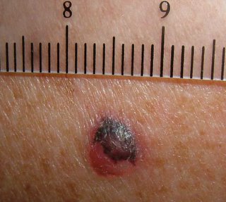
An excisional biopsy was performed.
Presumptive diagnosis is melanoma, probably nodular type.
Pigmented basal cell is another possibility, but rapid growth favors melanoma.
I do not think metastatic renal cell carcinoma would be pigmented.
January 25, 2006 -- Update
The biopsy showed a superficial spreading melanoma, 2.04 mm thick, Level IV.
The patient underwent a wide local excision with 2 cm margins. He elected not to have sentinal node biopsy.


0 comments:
Post a Comment