The patient is an 88 yo woman who came as a "walk-in" today.
She said she had a "volcano erupt" on her leg two weeks ago.
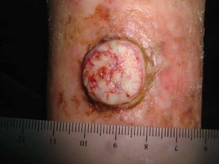
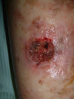
The examination shows an alert actve 88 yo woman with Type II skin. She has a dome-shaped tumor on her right shin which measures 2.1 cm in diameter. It seems to have a collarette surrounding it. She has marked actinic damage.

The history suggests keratoacanthoma. Some dermatologists call this "Squamous Cell Carcinoma -- Keratoacanthoma type." I suspect this is a coding ploy to make them seem malignant.
My question is what to do?
1) It would be hard to close after excision in this site.
2) If it is curetted and dessicated, it make take weeks to months to heal.
3) It could be observed, however, some of these larger lesions can be locally agressive.
4) Intralesional methotrexate is a possibility.
My instinct is to treat with C and E, but I'd like some suggestions. I might follow that with Aldara. I am open to suggestions.
Thank you.
David Elpern
May 4, 2006
At the advice of a few of you, I did a shave biopsy and curetted and dessicated the lesion. The base was mostly gritty (a good sign). I will follow closely. She's 88 year-old. So, I'll observe before any further intervention. May try imiquimod. Will play by ear.

She said she had a "volcano erupt" on her leg two weeks ago.
The examination shows an alert actve 88 yo woman with Type II skin. She has a dome-shaped tumor on her right shin which measures 2.1 cm in diameter. It seems to have a collarette surrounding it. She has marked actinic damage.

The history suggests keratoacanthoma. Some dermatologists call this "Squamous Cell Carcinoma -- Keratoacanthoma type." I suspect this is a coding ploy to make them seem malignant.
My question is what to do?
1) It would be hard to close after excision in this site.
2) If it is curetted and dessicated, it make take weeks to months to heal.
3) It could be observed, however, some of these larger lesions can be locally agressive.
4) Intralesional methotrexate is a possibility.
My instinct is to treat with C and E, but I'd like some suggestions. I might follow that with Aldara. I am open to suggestions.
Thank you.
David Elpern
May 4, 2006
At the advice of a few of you, I did a shave biopsy and curetted and dessicated the lesion. The base was mostly gritty (a good sign). I will follow closely. She's 88 year-old. So, I'll observe before any further intervention. May try imiquimod. Will play by ear.


