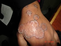
Abstract: 24 yo woman with 2 month history of transient plaques torso and extremities.
HPI: This 24 yo woman was diagnosed with hyperthyroidism in October of 2007. She was treated with radioactive iodine and carbamazole in November and December. Her dermatological manifestations began after both treatments. She reports ~ 20 episodes of painful plaques on torso and extremities. These last 1 - 3 days and clear completely. They are hot, tender, and painful in certain locations. She was first seen in my office on February 26, 2008 with an acute episode which was 24 hours old.
O/E: Healthy-appearing young woman. A solitary plaque was noted on the upper back. The borders were well-defined. The area was hot and painful and slightly erythematous. The patient had trouble taking her shirt off for the exam.
Photos:
Note: The border is outlined for clarity with a blue marking pen in photos 2 and 3.


Lab: She has had various blood tests done by other physicians and I've called for results. I ordered a CBC and ESR yesterday. Thyroid antibodies will be obtained unless her other physicians have ordered these.
Pathology: A deep incisional wedge biopsy into the panniculus was obtained.
Diagnosis: I have not seen anything like this. The short duration of the lesions suggests angioedema or urticarial vasculitis. But, I have never seen a similar case with such large lesions. One wonders about the relationship of her thyroid disease and possible autoantibodies.
Reason Presented and Questions: It is instructive to present an undiagnosed case for discussion. Others may have seen a similar patient. Every day, we see something unique to us. In some cases, our colleagues may be of invaluable assistance. Your comments are most welcome.
HPI: This 24 yo woman was diagnosed with hyperthyroidism in October of 2007. She was treated with radioactive iodine and carbamazole in November and December. Her dermatological manifestations began after both treatments. She reports ~ 20 episodes of painful plaques on torso and extremities. These last 1 - 3 days and clear completely. They are hot, tender, and painful in certain locations. She was first seen in my office on February 26, 2008 with an acute episode which was 24 hours old.
O/E: Healthy-appearing young woman. A solitary plaque was noted on the upper back. The borders were well-defined. The area was hot and painful and slightly erythematous. The patient had trouble taking her shirt off for the exam.
Photos:
Note: The border is outlined for clarity with a blue marking pen in photos 2 and 3.


Lab: She has had various blood tests done by other physicians and I've called for results. I ordered a CBC and ESR yesterday. Thyroid antibodies will be obtained unless her other physicians have ordered these.
Pathology: A deep incisional wedge biopsy into the panniculus was obtained.
Diagnosis: I have not seen anything like this. The short duration of the lesions suggests angioedema or urticarial vasculitis. But, I have never seen a similar case with such large lesions. One wonders about the relationship of her thyroid disease and possible autoantibodies.
Reason Presented and Questions: It is instructive to present an undiagnosed case for discussion. Others may have seen a similar patient. Every day, we see something unique to us. In some cases, our colleagues may be of invaluable assistance. Your comments are most welcome.


