This is default featured slide 1 title

Go to Blogger edit html and find these sentences.Now replace these sentences with your own descriptions.

This is default featured slide 2 title

Go to Blogger edit html and find these sentences.Now replace these sentences with your own descriptions.

This is default featured slide 3 title

Go to Blogger edit html and find these sentences.Now replace these sentences with your own descriptions.

This is default featured slide 4 title

Go to Blogger edit html and find these sentences.Now replace these sentences with your own descriptions.

This is default featured slide 5 title

Go to Blogger edit html and find these sentences.Now replace these sentences with your own descriptions.

Saturday, February 4, 2006

Nail Dystrophy

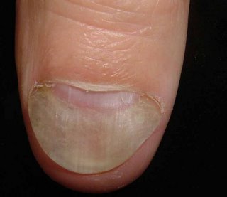
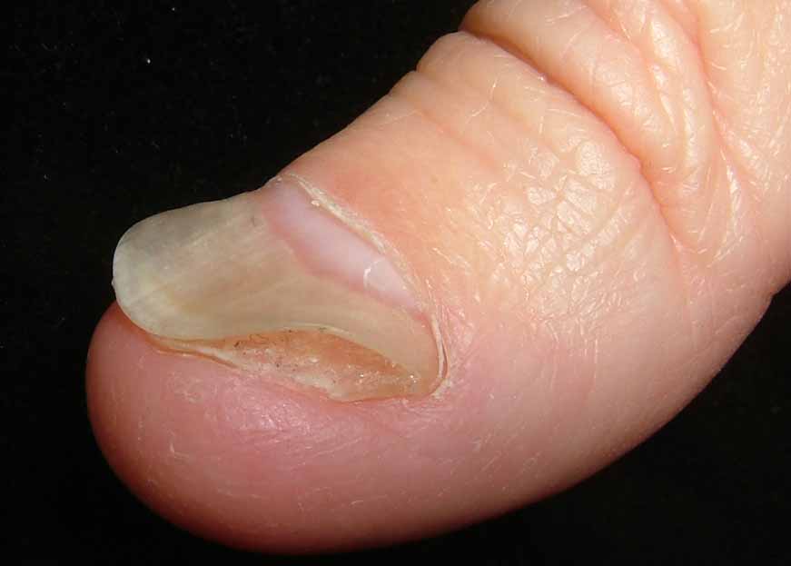
This 50 yo man has a three year history of a nail dystrophy.
I asked him to write out a detailed history:

"Started 18 months ago. Might well have contracted the condition while handling and cutting blocks of cheese as part of my former job. I used to handle and work with dozens of varieties of often moldy cheeses for three hours per week.

The condition began with a raised red ridge near the thumbnail. It was a connected series of raised red lumps about 1 to 2cm long and 3 or 4mm wide. This condition traveled progressively up and around my thumb. It always progressed one way up my thumb with the old area healing. It became a migrating „"ing of red". This went on for about six months. Often complete healing would occur, but then it would come up again right where it had left off and progress some distance more. Eventually it completely died out a little ways into the palm. There has been no trace of this symptom for a year now.

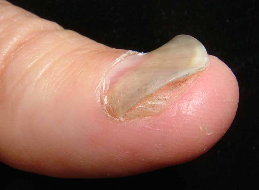
But starting also about 18 months ago the thumbnail became discolored (white and yellowish) and detached from the skin underneath. I've cut the thumbnail all the way back to the base in the hopes it will grow out correctly, but it remains detached from the skin underneath. Also the nail is misshapen: it is raised in some places and sunken in the middle. This has been the growth pattern for more than a year now. The skin underneath is clean but has a slight coating of what seems to be nail tissue on it, thus making it harder for the nail to attach itself. The nail is sound, just wavy and not attached underneath; because it is unattached underneath it is not pink, but rather it is white.


In the dermatology office there have been 2 negative KOH preps and a negative fungal culture. I did see some hyphae on one occasion. Ths question is: Is this a variant of funal nail disease, or are we dealing with onycholysis alone.

Your thoughts are appreciated.

Monday, January 30, 2006

Difficult BCC

On January 30, 2006, this 49 yo woman presented with a somewhat sclerotic plaque on the blub of the nose. By history, this has been present for 1.5 years. A 2 mm punch biopsy was taken at the site marked X.


If this turns out to be BCC, I will refer for micrographic surgery. Does anyone feel radiotherapy is appropriate for a patient of this age?

First Biopsy showed no evedence of BCC or malignancy. Repeat biopsy taken. Here is another clinical photo:

Thursday, January 26, 2006

Oral Hyperpigmentation

This 21 years student noticed hyperpigmentaiton of her buccal mucosae for one weeks.
She has gingival hyperpigmentation for years and has a family history of the latter.
Her health is good, and her only medication are albuterol and cromolyn sodium for asma.
No history of cancer in any first degree relation.


The examination show hyperpigmented buccal maculas measuring up to > 1 cm in diameter.
Gingival hyperpigmentation is present as well.


In the differential diagnosis I would consider pigmented fixed drug eruption, amalgam tattoo, melanoacanthoma. Biopsy may be offered. Melanoma is unlikely.

Presented by
Mordechai Pepe Grosbartt
Concepción, Chile

Tuesday, January 24, 2006

9 year old child with inflammatory alopecia

The patient is a 9 year-old girl who was in her usual state of health until September 5, 2005 when her mother applied a "hair relaxer" to her scalp. Within a few days, she developed inflammation. The process has persisted and over the past few months she has developed fluctuant areas over the scalp with alopecia and some scarring. There is moderate discomfort.

The examination shows a calm 9 year-old. Her scalp is involved globally with fluctuant nodules, some crusted. There are diffuse areas of alopecia. Some areas look scarred. Wood's light is negative. Posterior cervical lymph notes are enlarged and tender.


A bacterial and fungal culture were taken. Scalp biopsy will be performed and the patient will be started on prednisone 1 mg/kg/day and griseofulvin 25 mg/kg/day pending test results.

Working diagnosis is Inflammatory tinea capitis vs. dissecting cellulitis of the scalp.

Update February 9, 2006
The biopsies taken showed no fungfal elements, so the griseofulvin was discontinuted on Feb. 2, 2006 and patient was placed on cephalexin 50 mg/kg/day in divided doses. She did well.

On day 14, the fungal culture was positive and she was placed back on griseofulvin -- 25 mg/kg/day/ Her prednisone dose is 1 mg/kg/day.


Lesson: Scalp biopsy is not an absolute. Fungal culture takes ~ 2 weeks to be positive.
Dx: Inflammatory tinea capitis (the first diagnosis). Negative scalp bx threw me off.

Your comments are welcome.


Reference:
Tinea capitis mimicking dissecting cellulitis: a distinct variant.
Twersky JM, Sheth AP.
Int J Dermatol. 2005 May;44(5):412-4.
BACKGROUND: Tinea capitis is a common scalp dermatosis with several clinical
patterns. Only two patients with a presentation of tinea capitis mimicking
dissecting cellulitis have been described in the English literature.
OBSERVATION: We report a patient with tinea capitis mimicking dissecting
cellulitis who did not respond to griseofulvin therapy at 16 mg/kg/day but
eventually cleared after a protracted course of higher dose griseofulvin.
CONCLUSION: recognition of a dissecting cellulitis-like pattern of tinea capitis
will increase clinical suspicion and avoid inappropriate management of a
recalcitrant "dissecting cellulitis" in favor of prompt antifungal therapy of
appropriate dosage and duration for patients with this unusual variant of tinea
capitis.

Tuesday, January 10, 2006

Rapidly Growing Tumor

This 70 yo man presented on January 9, 2006 with a three to four month history of a lesion on his upper back. It was noticed by his wife. He has type III skin and a past history of renal cell carcinoma.


An excisional biopsy was performed.

Presumptive diagnosis is melanoma, probably nodular type.
Pigmented basal cell is another possibility, but rapid growth favors melanoma.
I do not think metastatic renal cell carcinoma would be pigmented.

January 25, 2006 -- Update
The biopsy showed a superficial spreading melanoma, 2.04 mm thick, Level IV.
The patient underwent a wide local excision with 2 cm margins. He elected not to have sentinal node biopsy.

Sunday, January 8, 2006

Suggestions for Wart Removal?

Dear Anak VGRD Members,

I am a G.P. trainee and have been having a hard time with a wart removal on a 3 year old's fifth toe. The difficulty is getting him to let anyone near it. I've seen him once, late in the day, and my preceptor asked that the child return for freezing with the help of sedation. We are now working on what to use. I prefer to avoid having to sedate the child, and have suggested duct tape in the interim (six days on, one day off with soaking). What suggestions or tricks would you have for treating a wart in this location in a three year-old? Thank you, Tammy Grimely, Hobart, Tasmania

Wednesday, January 4, 2006

Explosive Rosacea?

The patient is a 33 year-old executive V.P. who presents with a 6 month history of dramatic facial eruption. He had acne as a teenager but has been clear for many years. His health is good and he takes no medication by mouth. He has never used topical corticosteroids for his face.


The examination shows erythematous papules and small cysts on forehead, cheeks and chin. There are a few papules on the back, but the chest is clear.


Diagnosis: Florid Rosaca

Plan:
I will place on prednisone 20 mg bid for a week or so and isotretinoin 0.25 mg per kg for a cple of weeks and then increase to 0.5 mg per kg fir a month or two. Slowly incvrease isotretinolin to 1 mg per kg over 8 weeks and anticipate a six moth course. The prednisone will be tapered over a month to 6 weeks to avoid a flare.

I could have started with doxycycline; but isotretinoin may be more definitive; I welcom your thoughts.

Thank you,

David