This is default featured slide 1 title

Go to Blogger edit html and find these sentences.Now replace these sentences with your own descriptions.

This is default featured slide 2 title

Go to Blogger edit html and find these sentences.Now replace these sentences with your own descriptions.

This is default featured slide 3 title

Go to Blogger edit html and find these sentences.Now replace these sentences with your own descriptions.

This is default featured slide 4 title

Go to Blogger edit html and find these sentences.Now replace these sentences with your own descriptions.

This is default featured slide 5 title

Go to Blogger edit html and find these sentences.Now replace these sentences with your own descriptions.

Wednesday, March 19, 2008

Double Helix

We often see problems for which there may be no simple solution. Ear lobe keloids are encountered with regularity; but keloids of the helix and triangular fossae are unusual. Some of you may have a simple trick for patients like these:

Patient # 1.
Abstract: 25 yo woman with ear keloid.

HPI: This 25 yo Asian woman pierced the triangular fossa of her right ear 2 years ago and developed a keloid which is pruritic and whose appearance bothers her.

O/E:


Patient # 2
As I was getting case # 1 ready to publish on this site, a second patient presented for evaluation and treatment.
This is a 16 yo girl with a one year history of a keloid of the left triangular fossa. She had a professional piercing done two years ago. This lesion is painful.


This patient had an "Industrial Piercing" with a 14 guage stainless steel rod.

Comment: Earlobe keloids are commonly seen and reported. But I could find no helpful articles about helix and triangular fossa keloids. I suspect that these lesions are not rare, since I have seen two in a few weeks in a small New England town. Perhaps, these are harbingers of an epidemic! One of these young women pierced her own ear, and the other was a professional job.

Questions:
These can not be simply excised and then injected with TAC like the more common ear lobe keloid. Wound closure would be problematic.
How would you approach these women?
Any role for shave excision followed by imiquimod?
Do you think TAC alone will work? 20 mg per cc, 40 mg per cc?
Does anyone have experience with similar lesions?

Friday, March 14, 2008

Weird Erythema

Presented by Omid Zargari
Rasht, Iran

This patient referred to me with a weird erythema on his frontal area for the past two years.
He is a 45 y.o. kidney transplant patient whose medications include cellcept, cyclosporine, erythropoetin alpha and simvastatin. He believes that this began after he started simvastatin.


Your thoughts will be appreciated.

Monday, March 10, 2008

Acrolentiginous Melanoma from Malaysia

Here is a case from Jahor Baru, Malaysia. In North America, Europe and Australia the patient would have had more extensive studies; but one must keep in mind that there are disorders in Malaysia that are better handled there than here. Melanoma is far less common in SE Asia and their resources are allocated differently.

Abstract:
70 yo woman with ALM right sole

Presented by S.E. Choon, Consultant Dermatololgist

HPI: This 70 year-old Chinese lady presented in April 2006 with a 1-year history of a growth on right sole which was biopsied and diagnosed as melanoma. We do not have level and thickness. She refused below knee amputation and hence was referred to us in July 2006. She had radiotherapy in Sep/Oct 2006 in private centre.

P.M.H: Diabetes mellitus of 10 years, well-controlled with metformin 500 mg bd and glibenclamide 5mg daily.

O/E: see figure Fig. This shows her lesion in 2006 before XRT. There were no other positive findings. The current lesion very similar. Patient coming back next week.


Work UP:
CXR and CT neck, thorax and abdomen were clear in 2006 - in February 2008 CT scan last week showed several small right obturator lymph nodes. I was not too happy without a repeat CT and get one done last week. Size of LN not mentioned.

Pathology:
2006: Superficial spreading type (apparently down to subcutis for initial biopsy. Pathologist in Singapore wrote: "nodular type with vertical growth and ?superficial spreading"
2008: Recent lesion biopsy report was short and sweet-recurrent melanoma

Diagnosis: Acrolentigious Melanoma

Questions:
1. How would you classify this patient?
2. What would you recommend for this patient?
3. The patient initially refused amputation. Is there a lesser procedure that might help her now?

Your answers and comments are welcome.

Wednesday, February 27, 2008

Painful Edematous Plaques

Abstract: 24 yo woman with 2 month history of transient plaques torso and extremities.
HPI: This 24 yo woman was diagnosed with hyperthyroidism in October of 2007. She was treated with radioactive iodine and carbamazole in November and December. Her dermatological manifestations began after both treatments. She reports ~ 20 episodes of painful plaques on torso and extremities. These last 1 - 3 days and clear completely. They are hot, tender, and painful in certain locations. She was first seen in my office on February 26, 2008 with an acute episode which was 24 hours old.
O/E: Healthy-appearing young woman. A solitary plaque was noted on the upper back. The borders were well-defined. The area was hot and painful and slightly erythematous. The patient had trouble taking her shirt off for the exam.
Photos:
Note: The border is outlined for clarity with a blue marking pen in photos 2 and 3.

Lab:
She has had various blood tests done by other physicians and I've called for results. I ordered a CBC and ESR yesterday. Thyroid antibodies will be obtained unless her other physicians have ordered these.
Pathology: A deep incisional wedge biopsy into the panniculus was obtained.
Diagnosis: I have not seen anything like this. The short duration of the lesions suggests angioedema or urticarial vasculitis. But, I have never seen a similar case with such large lesions. One wonders about the relationship of her thyroid disease and possible autoantibodies.
Reason Presented and Questions: It is instructive to present an undiagnosed case for discussion. Others may have seen a similar patient. Every day, we see something unique to us. In some cases, our colleagues may be of invaluable assistance. Your comments are most welcome.

Wednesday, February 20, 2008

Facial Pigmentation in a 31 yr old woman with Protein S deficiency

A 31 yr old woman presented with one year history of pigmentation on her face. It appeared as sun burn and confined to the facial areas with sparing of the areas underneath the eyes and nose. It was made worse after application of some skin care products. She was otherwise well.

She had a past history of hypercoagulation and mitral valve prolapse with Protein S deficiency and is on warfarin.

Examination of the skin showed diffuse hyperpigmentation on the face extending to the frontal hairline, preauricular hairline and mentum. The areas under the eyes and nose were spared.

Blood counts and biochemistry were normal ANA serology mildly positive. Titre 1:80 (RR<80)

Differentials: photodermatitis > LE > melasma > irritant dermatitis

Q Is the serology titre of 1:80 significant? Could this be LE or melasma? Would you biopsy her skin? If you biopsy, where would you biopsy her? Thanks for your comments.

Sunday, February 10, 2008

Asymptomatic annular lesions on face and hands

Submitted by Khalil Alhamdi M.D.
Associate Professor of Dermatology
Basrah, Iraq

Abstract: A 25-year old woman with 3-year history of asymptomatic annular lesions on face and hands

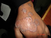
History: A 25-year-old woman presented with 3-year history of multiple asymptomatic annular lesions involving the face and the dorsa of both hands that gradually increase in size. She had received different modalities of treatment without improvement.

O/E: Young aged women presnted with multiple asymptomatic annular atrophied hyperpigmented patches with hyperkeratotic border that affect the face in a mask-shaped destribution and the dorsa of both hands.

Clinical Photo:


Lab: All relevant investigations were normal.

HPE: revealed features suggestive of porokeratosis


Diagnosis: Porokeratosis of Mibelli

Comments: This woman was misdiagnosed as fungal infection and lichen planus for which she received treatment without benefit. On clinical and histopathological bases we put her on topical 5Fu in addition to irregular courses of isotretnion because of poor compliance and inavailability of the latter drug in our country.

Questions:
1. What is the experience of our colleagues in seeing such unusual presentation?
2. What is your treatment suggestion to help this poor lady.

Friday, February 8, 2008

Umbilical Erosions

[ See end for final diagnosis]
Abstract:
71 yo woman with three week history of genital, anal and umbilical erosions.
HPI: This healthy 71 yo woman had vaginal prutitus for a few weeks. She saw her gynecologist who prescribed an estrogen cream. It got worse. She was then given clobetasol oint. It did not improve. She tried acyclovir ointment -- not much change. I saw her at this point. I recommended continuing clobetasol ointment, but after a few days getting worse. No new meds. Takes occasional acetoaminophen and diphenylhydramine.
O/E: Periumbilical erosive dermatitis. No frank vesicles. There was only faint erythema of the vulva and anal areas and very slight erosion left groin.


Lab: CBC normal, Chemistries normal. KOH from umbilicus negative. Bacterial culture taken.
Pathology: Biopsies for H&E and perilesional for DIF done Feb. 8, 2008
Diagnosis: I am considering the following:
A vesiculobullous disorder
Fixed drug eruption (but have no likely candidates)
Contact dermatitis unlikely.
HSV a long shot.
Periumbilical cellulitis? B-Strep perianal cellulitis can look similar
What have I missed?
Questions: What are your thoughts? Biopsy and culture should be ready in three days.

The bacterial skin culture grew out Group A Beta Strep. The pathology was consistent with cellulitis. No evidence of an acantholytic process. It is likely that this began with a perianal/vaginal streptococcal cellulitis and spread to the umbilicus. Periumbilical streptococcal cellulitis has not been reported in adults. The patient was started on Pen VK 250 mg qid and mupirocin ointment. Fout days later she was almost completely clear. Unfortunately, the fluorescent correction was not on when picture was taken.