This is default featured slide 1 title

Go to Blogger edit html and find these sentences.Now replace these sentences with your own descriptions.

This is default featured slide 2 title

Go to Blogger edit html and find these sentences.Now replace these sentences with your own descriptions.

This is default featured slide 3 title

Go to Blogger edit html and find these sentences.Now replace these sentences with your own descriptions.

This is default featured slide 4 title

Go to Blogger edit html and find these sentences.Now replace these sentences with your own descriptions.

This is default featured slide 5 title

Go to Blogger edit html and find these sentences.Now replace these sentences with your own descriptions.

Sunday, September 24, 2006

30 year old woman with changing mole


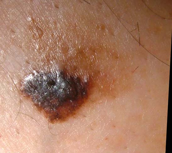
This 30 yo woman has noticed an enlarging pigmented lesion in her right axilla for almost a year. Her mother, a registered nurse, asked her to see a dermatologist. Her husband did not notice it.
The patient has Type II skin. The general cutaneous exam was unremarkable save for a 1.2 cm in diameter barely elevated plaque. There is a play of pigment and outline is irregular.

An excisional biopsy was performed.

Pathology: Malignant melanoma in situ , superficial spreading type. (Read by H. Byers of BU Skin Path who took photomicrographs) "The specimen exhibits a marked nested and lentiginous melanocytic proliferation with large severely atypical epithelioid cells. There is irregular nesting, focal confluent lentiginous melanocytic proliferation and cellular dyshesion."


She is scheduled for a wider excision, 0.5 cm on either side. No further work-up other than regular follow-up visits.

Your thoughts are appreciated.

Tuesday, September 19, 2006

Positive Band Aid Sign

Most dermatologists know about the "positive band aid sign." To me, it means a skin cancer has been lurking there for a while.

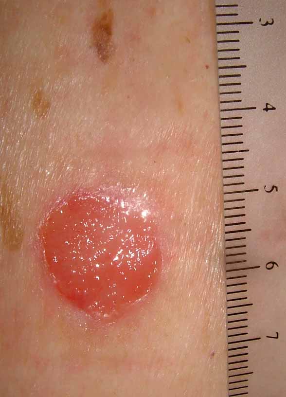
This 82 yo man came in with a one year history of a "sore" on his back. The drainage stained his shirt. When the band aid was removed, there was a one cm in diameter clean friable tumor, most likely a basal cell.

The lesion was shave biopsied and desicated and curetted. I'll affix a copy of the biopsy report when I get it.

How many of you use the term "Positive band Aid Sign?"

Wednesday, August 30, 2006

BCC of Eyelid

This 43 yo man has a one year history of a lesion of the left lower lid. It measures 8 mm in diameter.
Biopsy from "X" confirms "nodular BCC."
Question: What is best therapy?
1) Mohs
2) Mohs + closure by ophthalmic plastic surgeon
3) Ophthalmic plastic surgeon handle all
4) Another approach

Cada vendador allabe sus agujas.
Every peddler praises his needles.

Wednesday, August 2, 2006

Therapeutic Question

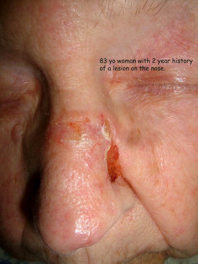
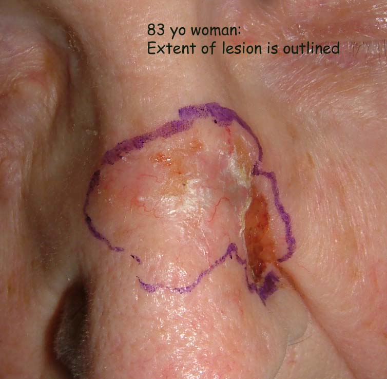
The patient is an 83 year-old woman with a two year history of a lesion on the nose.
Clinically, this is a basal cell. My question is what you would recommend as therapy and why?

1) Micrographic surgery with forehead advancement flap.*
2) Micrographic surgery with graft or allowed to heal by secondary intention.
3) Radiotherapy
3) Other


* Repair of defects on nasal sebaceous skin.
Dzubow LM.
Department of Dermatology, University of Pennsylvania Health System, Philadelphia, 19085, USA. leonarddzubow@comcast.net
BACKGROUND: Reconstructive procedures performed on sebaceous nasal skin are prone to partial flap necrosis, scar spread and inversion, and tissue mismatch. An ideal repair would optimize vascular integrity, minimize closure tension, and use adjacent tissue. OBJECTIVE: The purpose of this article is to describe a flap design and dynamics that permit satisfactory reconstruction of small- to medium-sized defects on nasal sebaceous tissue. METHODS: A modified advancement flap is described that may be used on central and off-midline defects of the nasal tip. RESULTS: Use of the modified advancement flap resulted in good cosmetic results with few adverse postsurgical events. CONCLUSIONS: The modified advancement flap satisfies the requirements of a hardy blood supply, minimization of closure tension, and use of adjacent tissue. The surgical results are predictable and rarely associated with complications.

Wednesday, July 26, 2006

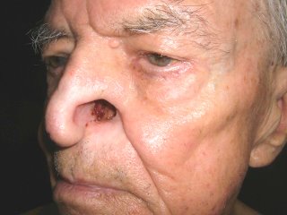
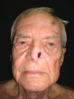
An Incidental Finding


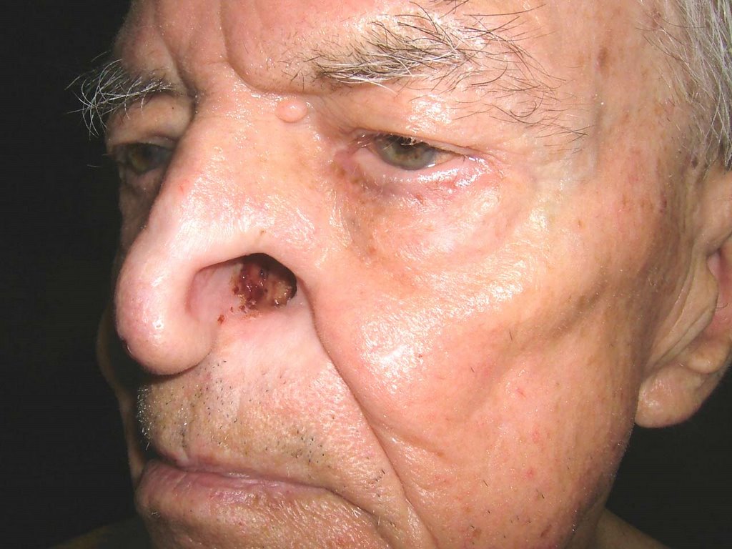
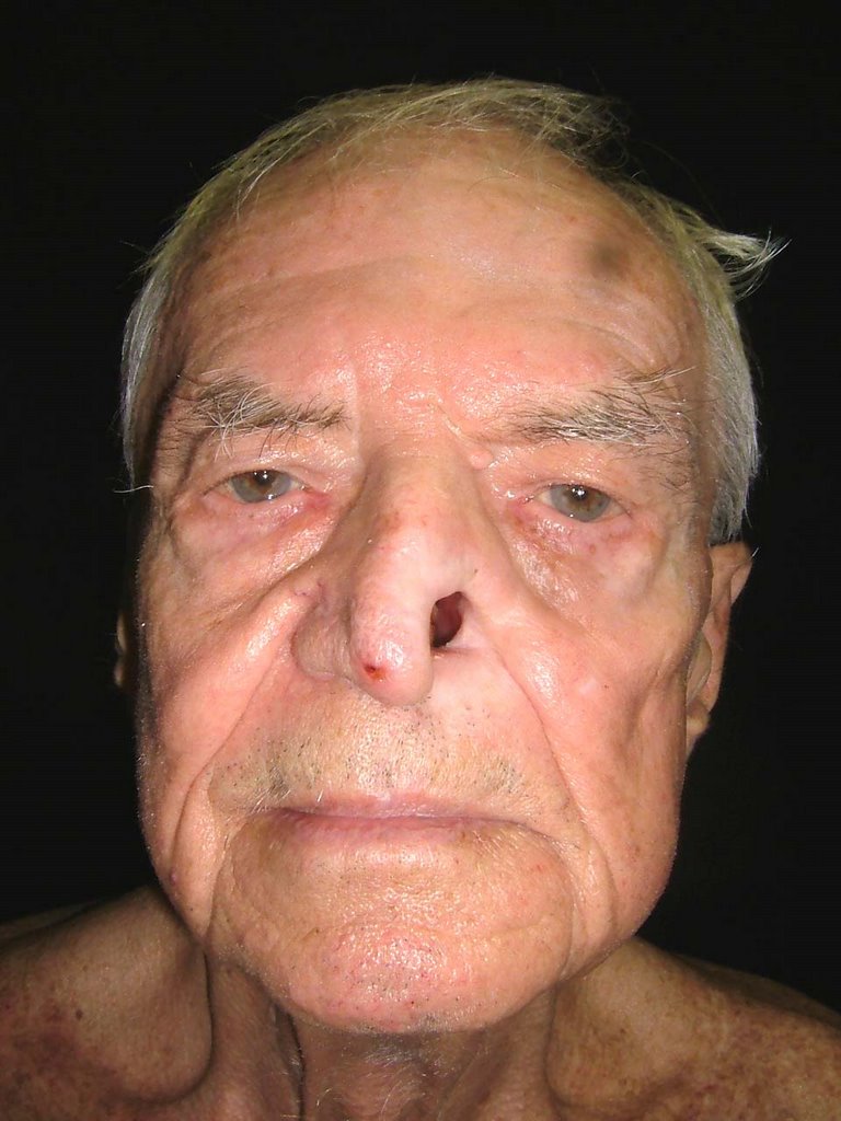
The patient is an 85 yo man who presented for generalized pruritus which has been present for two years. He has a history of prostate cancer and gastrointestinal bleeding. He's had a few transfusions over the past year or so and was concerned that they caused his itching.

He did not complain about his nose. I asked about it and he and his daughter gave this history: Three years ago he was hospitalized for pneumonia. He delvelped an inflammation of the ala nasi and was told it was "shingles." Biopsies were done and ruled out cancer. The left ala nasi was destroyed and this is how he healed.

Discussion. I wonder whether nasal oxygen was used and if the canula caused damage to the nose. It cold also have followed H. zoster. This is the picture that can be seen with Trigeminal Trophic Syndrome, a rare disorder that is thought to be traumatic in origin. Has anyone seen a similar picture and what are your thoughts.?

This is a good abstract:
Laryngoscope. 1988 Dec;98(12):1330-3.

Trigeminal trophic syndrome.

Arasi R, McKay M, Grist WJ.

Department of Dermatology, Emory University School of Medicine, Atlanta, GA.

Trigeminal trophic syndrome is an unusual condition also known as trigeminal
neurotrophic ulceration or trigeminal neuropathy with nasal ulceration. The
diagnosis is suggested when ulceration of the face, especially of the ala nasi,
occurs in a dermatome of the trigeminal nerve that has been rendered anesthetic
by a surgical or other process involving the trigeminal nerve or its central
sensory connections. A history of paresthesias and self-induced trauma to the
area further support the diagnosis. Neurological deficits causing trigeminal
trophic syndrome may result from surgical trigeminal ablation, vascular
disorders and infarction of the brainstem, acoustic neuroma, postencephalitic
parkinsonism, and syringobulbia. The following etiologies of nasal ulceration
should be excluded: postsurgical herpetic reactivation and ulceration, syphilis,
leishmaniasis, leprous trigeminal neuritis, yaws, blastomycosis,
paracoccidioidomycosis, lethal midline granuloma, pyoderma gangrenosum,
Wegener's granulomatosis, and basal cell carcinoma. In the case reported here,
the diagnosis of TTS was made primarily as a result of previous experience with
the syndrome, underscoring the importance of physician recognition of this
unusual disorder.

Monday, July 24, 2006

Tale of Two Lesions

I saw these two patients today. Your comments are most welcome. Path reports will be affixed when available.

1) 69 yo man with 1.5 month history of lesion right pinna. The lesion is slowly growing. The patient's son, a family doctor, asked him to make an appointment. This is a subtle lesion that could be a lentigo or a lentigo maligna. Two representative areas were biopsied.


2) A 55 year-old priest whose parishioner noted a lesion on his left arm and asked him to see a doctor about it. He's been aware of this lesion for 3 months. It appears that the darkly pigmented papule began in a lentigo. I favor a diagnosis of melanoma here but this could be a seborrheic keratosis. An excisional biopsy of raised and macular portions was performed and based on pathology I will recommend further therapy.

Wednesday, July 19, 2006

Hypopigmentation in atopic dermatitis

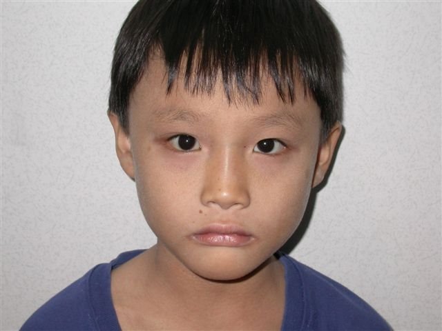
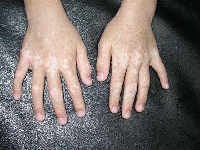
This is a 7-year-old boy who developed multiple patches of hypopigmentation on the dorsum of the hands for 3 years. He had a history of atopic dermatitis and had severe eczema of the hands prior to development of the hypopigmentation on the hands. According to the mother, he had secondary infection with crusts formation on the hands. The lesions were patchy hypopigmented macules mainly on the dorsum of the hands and feet. There were no family history of similar pigmentation. Wood's lamp examination however showed accentuation of the hypopigmented lesions.

Based on history this is most likely post inflammatory. Other differential considered includes vitiligo. Vitiligo is probably unlikely in this patient as the hypopigmentation developed after he had eczema with secondary infection of the hands and feet.

Any suggestions for this hypopigmentation? Would UVB or tacrolimus ointment help?

Thanks for your comments.