The Pathology Report: ATYPICAL LYMPHOID HYPEPLASIA
"Superficial and deep, nodular and diffuse lymphohistiocytic infiltrate forming follicular germinal centers consistent with atypical lymphoid hyperplasia.
NOTE : Immunostaining reveals a mixture of T-cells (CD3) and B-cells (CD-20). No clonal proliferation is seen on Kappa and Lambda staining. No CD-30 positive cells are noted. These findings are supportive of a lymphoid hyperplasia. If the clinical suspicion persists, follow-up of the patient is suggested"
Given the location, one wonders if this could be secondary to a tick bite (common in our area). See; Pediatr Dermatol. 2001 Nov-Dec;18(6):481-4.
Persistent atypical lymphocytic hyperplasia following tick bite in a child:
report of a case and review of the literature.
Hwong H, Jones D, Prieto VG, Schulz C, Duvic M.
Department of Internal Medicine Specialties, Section of Dermatology, University
of Texas-M.D. Anderson Cancer Center, Houston, Texas 77030, USA.
We report a 6-year-old girl who developed a red papule on the posterior neck at
the site of a previous tick bite. Initial biopsy was performed a year after the
bite and the specimen showed a dense lymphoid infiltrate with admixed CD30+
cells. The patient was referred to our center because of concern about the
development of a CD30+ lymphoproliferative disorder. The lesion was completely
excised. Histology showed no evidence of a clonal lymphoproliferative disorder
or Borrelia infection, but persistence of CD30+ cells. This case demonstrates
that a tick bite reaction can persist for more than 1 year and show
immunophenotypic and morphologic overlap with a CD30+ lymphoproliferative
disorder. Complete history with thorough clinical and histopathologic evaluation
is necessary to arrive at the correct diagnosis.
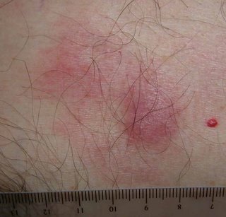
The patient is a 52 yo engineer who presents with a 2 month history of a 1.5 cm in diameter asymptomatic somewhat "spongy" presternal nodule surrounded on one side with macular non-blanchable erythema.
The clinical appearance is non-diagnostic. This may be an infiltrative process, possibly a malignancy. I have not seen anything like this before with the possible exception of a Merkel Cell carcinoma. Punch biopsies were taken from the nodule and the surrounding erythema.
The results should be back on April 3.
What are your thoughts?


0 comments:
Post a Comment