This is default featured slide 1 title

Go to Blogger edit html and find these sentences.Now replace these sentences with your own descriptions.

This is default featured slide 2 title

Go to Blogger edit html and find these sentences.Now replace these sentences with your own descriptions.

This is default featured slide 3 title

Go to Blogger edit html and find these sentences.Now replace these sentences with your own descriptions.

This is default featured slide 4 title

Go to Blogger edit html and find these sentences.Now replace these sentences with your own descriptions.

This is default featured slide 5 title

Go to Blogger edit html and find these sentences.Now replace these sentences with your own descriptions.

Thursday, May 4, 2006

Folliculitis and Hair Loss

The patient is a 30 year-old man with a 2 year history of folliculitis and mild hair loss. He has erythematous papules and a few pustules scattered over the scalp. Cultures have been negative. Biopsy showed only inflammation c/w folloculitis. Minocycline has been of marginal help, but process recurrs when he stops. He has an occasional papule on his cheeks but no signs of rosacea. His hair is globally thin.

Question: Is this folliculitis decalvans? Would isotretinoin be worth trying? He is also interested in hair transplantation.


Friday, April 28, 2006

Volcanic Eruption

The patient is an 88 yo woman who came as a "walk-in" today.
She said she had a "volcano erupt" on her leg two weeks ago.

The examination shows an alert actve 88 yo woman with Type II skin. She has a dome-shaped tumor on her right shin which measures 2.1 cm in diameter. It seems to have a collarette surrounding it. She has marked actinic damage.


The history suggests keratoacanthoma. Some dermatologists call this "Squamous Cell Carcinoma -- Keratoacanthoma type." I suspect this is a coding ploy to make them seem malignant.

My question is what to do?
1) It would be hard to close after excision in this site.
2) If it is curetted and dessicated, it make take weeks to months to heal.
3) It could be observed, however, some of these larger lesions can be locally agressive.
4) Intralesional methotrexate is a possibility.

My instinct is to treat with C and E, but I'd like some suggestions. I might follow that with Aldara. I am open to suggestions.

Thank you.

David Elpern

May 4, 2006
At the advice of a few of you, I did a shave biopsy and curetted and dessicated the lesion. The base was mostly gritty (a good sign). I will follow closely. She's 88 year-old. So, I'll observe before any further intervention. May try imiquimod. Will play by ear.

Saturday, April 22, 2006

Cream Complacency

A 60 yo man was seen yesterday for pruritic papules on scalp and extremities. He had seen another dermatologist over the years and had been treated with a host of topicals including liquid nitrogen. Biopsy showed Prurigo nodularis.


[Contents of Bag Brought in by Patient]


After a few years, he developed a febrile illness, was seen in the ER where a CBC showed a WBC of 80,000. He was eventually diagnosed with a rare T-cell lymphoma. Four courses of CHOP have put him in remission.

If his prurigo becomes more active again, one wonders if this will be a harbinger of recurrence.

Sobering lesson. Prurigo may be realated to an underlying malignancy. We can all be lulled into a cream complacency and miss an important clue. I can think of one or two patients over the years with unexplained excoriations who turned out to have a lymphoproliferative malignancy.

Reference:
Seeburger J, Anderson-Wilms N, Jacobs R.
Lennert's lymphoma presenting as prurigo nodularis.
Cutis. 1993 May;51(5):355-8.
Section of Dermatology, Loma Linda University School of Medicine, California.
Abstract:
Lennert's lymphoma is a peripheral T-cell lymphoma that only rarely involves the skin. We present the case of a forty-two-year-old man who experienced severe pruritus for ten months. He was repeatedly diagnosed as having neurodermatitis
and prurigo nodularis before subtle hematologic clues suggested, and subsequent examination of bone and lymph node biopsy specimens confirmed, the diagnosis of Lennert's lymphoma. This report describes the case and presents a brief review
of Lennert's lymphoma.

Friday, April 21, 2006

U.P in a Newborn


I saw a new born today at the neonatal ward. He is the first child, just delivered today by LSCS for fetal distress. Noted to have generalized erythematous wheals and papules. Some of the wheals and papules appear to form blisters. The mother had no history of any infection during her pregnancy. On examination the child appeared comfortable and not in distress. Sleeping and quiet. Afebrile. Generalised erythematous raised papules and wheals were noted on the face, trunk and upper limbs. Some of the wheals blanche with pressure. Tried to elicit darier’s sign – mild erythema but not raised. The wheals were intensely erythematous and inflamed on certain parts of the neck and face.

Clinically he has urticaria pigmentosa

His blood counts : TWBC 3100 Eosinophils 20% 
Platelets 44 000
G6PD - pending

Though the rash appeared intense, the child was comfortable. Would you have started him on oral hydroxizine? Would you investigate further – hematological malignancy, etc the parents were advised about trigger factors (rubbing) and drugs (anesthetics, etc). thanks, Henry Foong

Thursday, March 30, 2006

Unknown

What are your thoughts?


The Pathology Report: ATYPICAL LYMPHOID HYPEPLASIA
"Superficial and deep, nodular and diffuse lymphohistiocytic infiltrate forming follicular germinal centers consistent with atypical lymphoid hyperplasia.

NOTE : Immunostaining reveals a mixture of T-cells (CD3) and B-cells (CD-20). No clonal proliferation is seen on Kappa and Lambda staining. No CD-30 positive cells are noted. These findings are supportive of a lymphoid hyperplasia. If the clinical suspicion persists, follow-up of the patient is suggested"

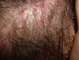
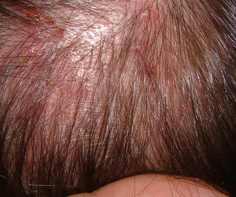
Given the location, one wonders if this could be secondary to a tick bite (common in our area). See; Pediatr Dermatol. 2001 Nov-Dec;18(6):481-4.

Persistent atypical lymphocytic hyperplasia following tick bite in a child:
report of a case and review of the literature.

Hwong H, Jones D, Prieto VG, Schulz C, Duvic M.

Department of Internal Medicine Specialties, Section of Dermatology, University
of Texas-M.D. Anderson Cancer Center, Houston, Texas 77030, USA.

We report a 6-year-old girl who developed a red papule on the posterior neck at
the site of a previous tick bite. Initial biopsy was performed a year after the
bite and the specimen showed a dense lymphoid infiltrate with admixed CD30+
cells. The patient was referred to our center because of concern about the
development of a CD30+ lymphoproliferative disorder. The lesion was completely
excised. Histology showed no evidence of a clonal lymphoproliferative disorder
or Borrelia infection, but persistence of CD30+ cells. This case demonstrates
that a tick bite reaction can persist for more than 1 year and show
immunophenotypic and morphologic overlap with a CD30+ lymphoproliferative
disorder. Complete history with thorough clinical and histopathologic evaluation
is necessary to arrive at the correct diagnosis.


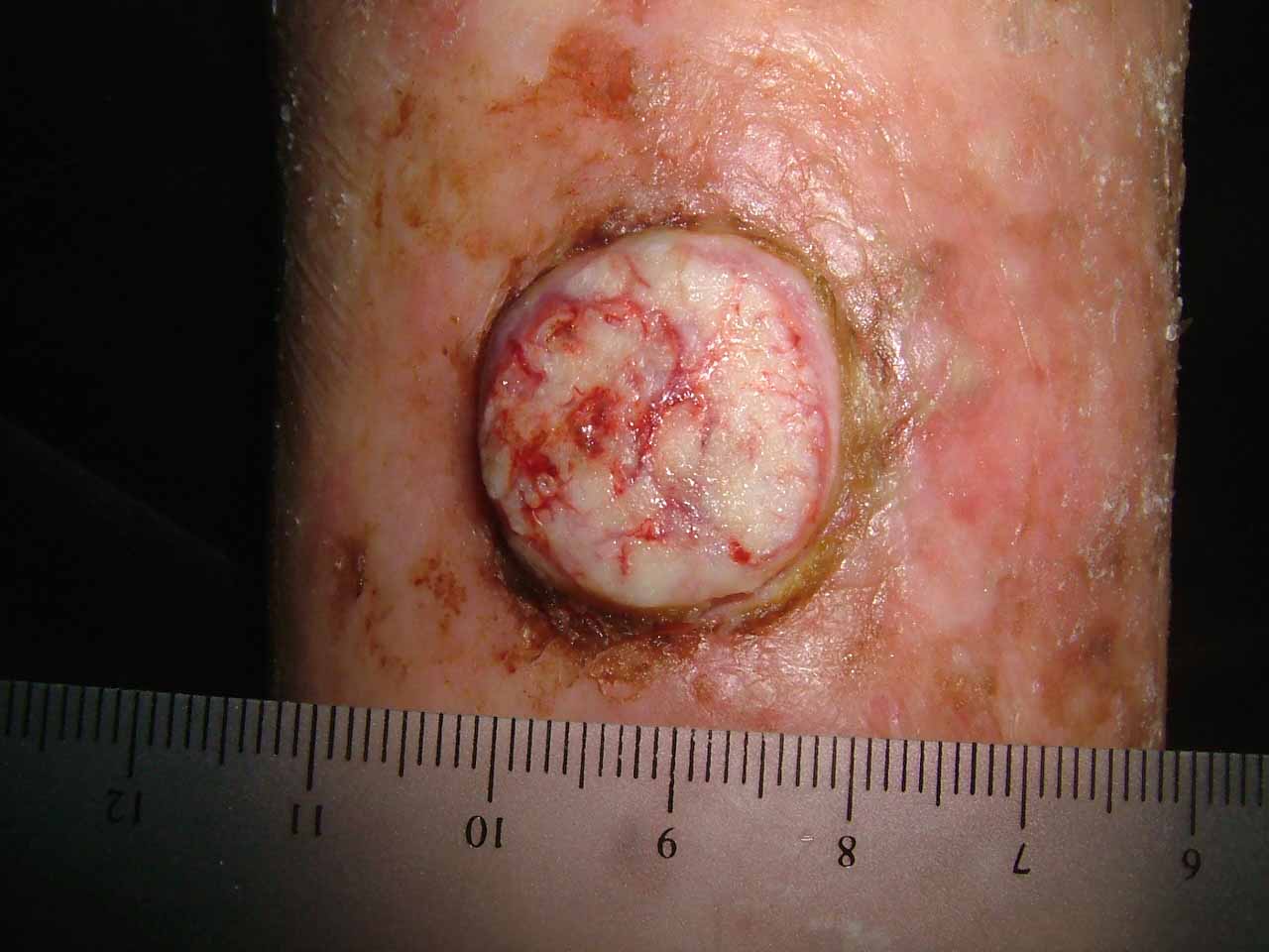
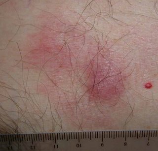
The patient is a 52 yo engineer who presents with a 2 month history of a 1.5 cm in diameter asymptomatic somewhat "spongy" presternal nodule surrounded on one side with macular non-blanchable erythema.
The clinical appearance is non-diagnostic. This may be an infiltrative process, possibly a malignancy. I have not seen anything like this before with the possible exception of a Merkel Cell carcinoma. Punch biopsies were taken from the nodule and the surrounding erythema.
The results should be back on April 3.


What are your thoughts?

Saturday, March 18, 2006

“NAILING DOWN A DIAGNOSIS”


Presented by
Brian T. Maurer, Enfield, Connecticut


D.S. is a 72-year-old left-handed male with a three-year history of periungual inflammation coupled with periodic cracking, separation and nail loss on the 3rd, 4th and 5th fingers of the left hand. The pads of these fingers also appear erythematous and swollen with dry cracked skin.

Initially, a local dermatologist prescribed topical Lamisil without effect. A subsequent month-long course of oral griseofulvin likewise did nothing. The periungual inflammation seemed to respond to topical erythromycin; the patient reports that all symptoms subsided over the summer months, only to return with the cold autumn weather.

An amateur gardener and golfer, D.S. wears a golf glove on his left hand during summer golfing season (when the condition seems to subside). He reports that his left hand usually feels colder than the right in winter time.

With the exception of one discolored nail of the left great toe, the nails of his remaining fingers and toes appear normal; he exhibits no other form of dermatitis.

Questions: Given the history and clinical course, what is the diagnosis? Suggestions for treatment?

Monday, March 13, 2006

Digital Tumor

This 61 yo man presented with a 30 year history of an annoying tumor of his right index finger. It interferes with his writing in that it abuts on the area in contact with a pen or pencil. He has pared it down on numerous occasions and it has been unsuccessfully treated with cryotherapy in the past. The clinical picture is that of an acquired digital fifrokeratoma (see reference below). An excisional biopsy is scheduled.


Dupre A, Christol B, Bories M.
[Acquired fibro-keratoma. Report of 8 cases]
Ann Dermatol Venereol. 1977 Oct;104(10):611-5. [Article in French]

The acquired fibro-keratoma is a benign fibro-epithelial tumor of acquired
nature. I was set apart as a distinct entity in 1968 by Bart et al. who named it
"acquired digital fibro-keratoma". It is predominantly, although not
exclusively, located on the fingers and toes, near the phalangeal joints. The
authors report 8 personal cases and make a short general review of the disease.
The lesion is slightly prominent, well defined and surrounded by a
hyperkeratotic collar. It protrudes on the surface of the skin much like a
hernia. Histologically, it is a dermo-epidermal tumor. There is a proliferation
of connective tissue under the papillomatous, hyperacanthotic and orthokeratotic
epidermis. The mature collagen bundles, voluminous fibroblasts and numerous
enlarged capillaries are parallel to the vertical axis of the tumor. The
acquired fibrokeratoma must be differentiated from other cutaneous tumors,
particularly fibromas and Koenen's tumors. The etiology is unknown. However, it
was found in some cases that a trauma had contributed to the development of the
tumor. The only possible treatment is surgical excision.