This is default featured slide 1 title

Go to Blogger edit html and find these sentences.Now replace these sentences with your own descriptions.

This is default featured slide 2 title

Go to Blogger edit html and find these sentences.Now replace these sentences with your own descriptions.

This is default featured slide 3 title

Go to Blogger edit html and find these sentences.Now replace these sentences with your own descriptions.

This is default featured slide 4 title

Go to Blogger edit html and find these sentences.Now replace these sentences with your own descriptions.

This is default featured slide 5 title

Go to Blogger edit html and find these sentences.Now replace these sentences with your own descriptions.

Wednesday, February 12, 2014

Prurigo Nodularis with KA Like Lesions


Abstract: 63 yo man with 10 month history of intense pruritus and excoriated papules and nodules

HPI:   This 63 yo retired radio announcer presents with a 10 month history of intense pruritus and excoriated papules and nodules. He is in reasonable health.  Medications include Welbutrin (bupropion) and occasional prednisone for his itching.  He's tried topical steroids and anti-histamines without relief.  Smokes ~ 5 cigarettes a day.

O/E: Skin thin from actinic damage.  There are excoriated papules and nodules on the torso and extremities.

Clinical Photos:


Lab:  CBC, chemistries normal.  IgE 867 IU/Ml

Pathology:  Initial bx signed out as SCC.  Since he has scores of lesions repeat biopsies of an early and more developed lesion were taken.

Diagnosis:

Discussion and Questions:

References:
1. Journal of the American Academy of Dermatology
Volume 69, Issue 3 , Pages 426-430, September 2013
Keratoacanthomas arising in association with prurigo nodules in pruritic, actinically damaged skin
Timothy P. Wu, BA, Kristen Miller, MD, David E. Cohen, MD, Jennifer A. Stein, MD, PhD  jennifer.Stein@nyumc.org

2. J Clin Pharm Ther. 2013 Feb;38(1):16-8. doi: 10.1111/jcpt.12005. Epub 2012 Sep 26.
Treatment of prurigo nodularis with pregabalin.
Mazza M, Guerriero G, Marano G, Janiri L, Bria P, Mazza S.

3. Dermatol Ther. 2010 Mar-Apr;23(2):194-8. doi: 10.1111/j.1529-8019.2010.01314.x.
Therapeutic hotline: Treatment of prurigo nodularis and lichen simplex chronicus with gabapentin.
Gencoglan G, Inanir I, Gunduz K.  (no real data on patient background)

4. Eur J Dermatol. 2008 Jan-Feb;18(1):85-6. Epub 2007 Dec 18.
Gabapentin for the treatment of recalcitrant chronic prurigo nodularis.
Dereli T, Karaca N, Inanir I, Oztürk G.  Available Free Full Text.

Saturday, February 1, 2014

Changing Nevus

Presented by:
Dr. Salvatore Donatello
Dermatologica e Venereologia. Catania, Sicilia


Abstract:  44 year old man with changing naevus

HPI:  The patient was seen in July of 2012 for a general cutaneous exam. He has Type IV skin. A naevus was noted on his right shoulder.  Dermatoscopic exam was felt to be not particularly worrisome but he was asked to return to have the lesion rechecked in six months.  He returned 18 months later.  The patient thought it had been present for many years and had not noted change.

O/E: July 2012:  5 mm in diameter dark brown papule  on right shoulder.  At the time, I thought the dermatoscopic exam looked normal.  It did look like an active lesions

January 2014:  The lesion is still 5 mm in diameter.  However, the brown clods noted on dermatoscopy in 2012 have disappeared and the lesion now looks uniformly dark gray.

Clinical photos:

Clinical Photo 31.1.14
Dermatoscopic Photo 31.1.14
Plan:  The lesion was excised with 3 mm margins on 31.1.14

Follow-up:  The biopsy report showed that this is an intradermal melanocytic without any atypical features. The brown clods in the first biopsy most likely are a sign of an active growing lesion.

Discussion:  Neither lesion on its own was very worrisome.  However, fortunately we have dermatoscopic images from July 2012 and January 2014.  The brown clods have disappeared and the lesion looks uniformly gray. Although the patient was asked to return in six months he did not do so.  We have no way of knowing if the change would have been noted then.
The pathology will tell us the story.  Is this an evolving naevus or a melanoma.  The practice of medicine can be humbling. There are few articles on evolving naevi in the literature.  This case will teach us something.

We will add an update after the biopsy is signed out. I have sent the specimen to a dermatologist in Napoli with a special interest in pigmented lesions.

Saturday, January 18, 2014

Periodic Shedding of the Nails


Abstract:  2 year old girl with seven month history of nail shedding

HPI:  This otherwise healthy 2 year-old girl has been losing nails since about 18 months of age.  By history, the nails turn black and then are shed.  The patient's father says his small toe nail sheds periodicaly and his father may have a nail dystrophy, also.  This grandfather has a muscular disorder.

OE:  There are six or seven nails with which shoe subungual hemorrhage, onycholysis, nail dystrophy or absent nails.  Fingers are more affected than toenails, however, many of the toenails are dystrophic.

Clinical Photos:
12/10/13


1/14/14


Here is a two week f/u of nail direcctly about this photo.  It's amazing how quickly the nails of young children grow.


Diagnosis:
The clinical picture is consistent with what has been called Periodic Shedding of the Nails (PPS).  There are only two articles on this entity and both are difficult to obtain.  It is possible that what was called PPS is really a localized variant of Epidermolysis Simplex. Since this is such a young child, the true nature of the disorder may become evident with the passage of time.

Discussion: We will try to obtain some opinions from experts who may have some experience here.  It is likely that this is autosomal dominant with varying degrees of penetrence.  It may improve with age.  Trauma probably plays a role in damaging the nail bed, so one wonders if there is some defect of the nail matrix or bed.  The finger pad erosion pictured above may indicate that the problem is more wide-spread than the nail matrix.


References
1. Cutis. 1980 Jun;25(6):622-3.
Familial dystrophic periodic shedding of the nails.
Martin S, Rudolph AH.
Abstract  A patient with an autosomal dominant nail dystrophy characterized by periodic, asymptomatic shedding of the nails followed by regrowth is described herein. This highly penetrant disorder is similar to two earlier cases found in the dermatologic literature.

2.  Br J Dermatol. 1973 May;88(5):497-8.
Periodic shedding of the nails.
Main RA.

3. Localized epidermolysis bullosa simplex (Weber-Cockayne type).
Villaseñor-Park J, English JC. J Pediatr Adolesc Gynecol. 2011 Dec;24(6):410-2.


Saturday, January 11, 2014

Desmoplastic Melanoma


Abstract: 83 yo man with a mixed desmoplastic melanoma of the scalp


HPI: In August 2013, this 83 yo man was seen with an 8 mm diameter nodule on the left parietal scalp present for  4 – 5 months and growing rapidly.  He had a history of two thin melanomas excised from the left forhead and left temple from 2009 – 2011.  In reviewing the biopsy reports, it can not be determined if they were both the same lesion or two separate tumors.

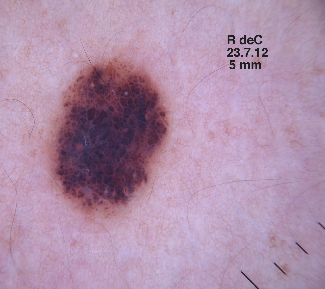
O/E: 8 mm pink to red well-circumscribed nodule left parietal. The dermatoscopic image shows multiple polymorphous blood vessels with central crystalline structures and a pink hue (vascular blush ) in the background.  No palpably enlarged lymph nodes.

Clinical Photos:


Dermoscopic Image - Courtesy of Yoon Cohen

Pathology:  Desmoplastic melanoma > 7.35 mm thick.  Level 5.  The tumor was/was not purely desmoplastic but over 10% of the cells has a spindle and epitheloid component as well.
Lab and Xray:
All blood studies including LDH within normal range.
P.E.T. Scan no abnormal foindings

Surgical Treatment: Patient underwent a WLE with 2 cm margins on October 2, 2013.  The defect was closed with a fasciocutaneous flap. Sentinel lymphnode biopsy was not performed.

Diagnosis: Desmoplastic melanoma with mixed histology, greater than 7.25 mm thick  No evidence of distant spread.

The patient has been seen at two centers for discussion of further treatment.  Local radiotherapy was recommended at both, although two different protocols were discussed.  The patient is undecided if he wants radiotherapy at this time.

Discussion:  The literature indicates that local radiotherapy reduces the incidence of local recurrence, but it is unclear if it provides an overall survival benefit.  At least one study suggests that patients undergoing local radiotherapy have a lower survival (ref 1 below).  It does not mention if these patients had more advanced disease that those who did not receive radiotherapy.

Questions:  What would your recommendations to this patient be?  Would you favor local radiotherapy?  See Discussion above.
References:
1. Desmoplastic melanoma - the step-child in the melanoma family?
Wasif N, Gray RJ, Pockaj BA.
J Surg Oncol. 2011 Feb;103(2):158-62.
Abstract
BACKGROUND AND OBJECTIVES: Desmoplastic melanoma (DM) is a rare variant of cutaneous melanoma. Our goal was to study the surgical management of DM, identify prognostic factors, and impact of treatment options.
METHODS: Patients with DM (n = 1,735) were identified from the Surveillance, Epidemiology, and End Results database (1988-2006).
RESULTS: The median age of the study population was 69 years and overall survival (OS) at 5 years 65%. DM was more common in males (65%), most commonly found on the head and neck (51%), and had a mean thickness of 2.97 mm. Patients undergoing a wide local excision (WLE; ≥1 cm) had improved 5-year OS compared to a simple excision (<1 cm) or biopsy alone (67% vs. 60% vs. 45%, respectively, P < 0.001). Of 505 patients (29%) undergoing sentinel node biopsy (SLNB), only 14 (2.8%) were positive. Traditional prognostic factors such as Breslow thickness, nodal positivity, and ulceration did not predict survival. On multivariate analysis only adjuvant radiation therapy [HR 1.65 (95% CI 1.17-2.31)] and WLE correlated with survival [HR 0.47 (95% CI 0.32-0.69)].
CONCLUSIONS: Desmoplastic melanoma does not share traditional prognostic factors with the melanoma family. Surgical resection with wide margins is needed to optimize survival and routine SLNB may be unnecessary. Furthermore, patients who received adjuvant radiation were at increased risk of dying (HR 1.65) and had decreased OS at 5 years (66% no radiation vs. 50% for adjuvant radiation, P < 0.001). Importantly, none of the traditional prognostic factors for cutaneous melanoma, such as site, Breslow thickness, Clark level, ulceration, and nodal status, had any impact on survival on univariate or multivariate analysis.
 
2. Mixed versus pure variants of desmoplastic melanoma: a genetic and immunohistochemical appraisal. Free Full Text
Miller DD, Emley A, Yang S, Richards JE, Lee JE, Deng A, Hoang MP, Mahalingam M.
Mod Pathol. 2012 Apr;25(4):505-15. Free Full Text
Abstract
Desmoplastic melanoma is subclassified into pure and mixed variants with a higher rate of lymph node metastasis in the latter. Given that reasons for these biological differences are not currently known, we investigated these subtypes with techniques that included genetic and immunohistochemical analyses of 43 cases of desmoplastic melanoma (24 pure, 19 mixed). Direct DNA sequencing was performed on BRAFV600E, RET gene (coding region on exon 11) and KIT (exons 11, 13 and 17). Immunohistochemical stains were performed with antibodies to markers of significance with respect to biological potential of nevomelanocytic proliferations and/or desmoplastic melanoma (Ki-67, CD117, nestin, clusterin, SOX10 and CD271/p75NTR). Polymorphism at the RET coding region (RETp) was noted in 33% of pure (8/24 cases) versus 24% of mixed (4/17 cases); BRAFV600E was absent in all cases of pure (0/24 cases) versus 6% of mixed (1/17 cases); no mutations were found in any of the cases on analyses of exons 11, 13 and 17 of the c-KIT gene (P=NS for all). For immunohistochemical analyses of pure versus mixed: mean percentage of Ki-67 nuclear positivity was 5% (s.d.=5.6) versus 28% (s.d.=12.6, P<0.001); CD117 stained 26% (6/23 cases) versus 78% (14/18 cases, P<0.01); nestin stained 83% (n=19/23 cases) versus 89% (16/18 cases, P=NS); clusterin stained 4% (1/23 cases) versus 6% (1/18 cases, P=NS); SOX10 87% (20/23 cases) versus 94% (17/18 cases, P=NS) and CD271 stained 61% (14/23 cases) versus 67% (12/18 cases, P=NS). Increased CD117 staining in the mixed variant suggests that alterations in the KIT protein may be involved in tumor progression. In addition, the proliferative index of the mixed variant was higher than that of the pure variant.

3.Subclassification of desmoplastic melanoma: pure and mixed variants have significantly different capacities for lymph node metastasis.
George E, McClain SE, Slingluff CL, Polissar NL, Patterson JW.
J Cutan Pathol. 2009 Apr;36(4):425-32.
Abstract
BACKGROUND: There is disagreement about the behavior and optimal management of desmoplastic melanoma (DM), particularly regarding the incidence of lymph node (LN) involvement. Recently, investigators have noted the frequently heterogeneous histologic composition of DM and have found significant differences between pure desmoplastic melanoma (PDM) (>or=90% comprised of histologically typical DM) and mixed desmoplastic melanoma (MDM) [>or=10% DM and >10% conventional melanoma (CM)].
METHOD: We reviewed 87 cases of DM comparing the histologic and clinical features of PDM (n = 44) to MDM (n = 43).
RESULTS: At surgical staging, there were LN metastases in 5 of 23 (22%) MDM patients, whereas all 17 PDM patients had negative LN biopsies (0%) (p = 0.04). PDM was less often clinically pigmented (36% vs. 67%) and had a lower mean mitotic index (1.3 vs. 3.0).
CONCLUSIONS: There are differences between PDM and MDM, the most important of which is the incidence of LN involvement. Our findings support the clinical utility of classifying DM into pure and mixed subtypes because the negligible rate of nodal involvement in PDM does not support the routine performance of sentinel LN biopsy in this subgroup of melanoma patients. In contrast, the incidence of LN involvement in MDM is comparable to that of CM.

4.  Desmoplastic malignant melanoma: a systematic review.
Lens MB, Newton-Bishop JA, Boon AP. Br J Dermatol. 2005 Apr;152(4):673-8.
Abstract
Prompt definitive surgical excision is the treatment of choice for DM. Improved knowledge of the clinical behaviour and histological features of DM is important for more effective management of patients with DM.

5. Desmoplastic melanoma: a review.
Chen LL, Jaimes N, Barker CA, Busam KJ, Marghoob AA.
Abstract:  Desmoplastic melanoma (DM) is a variant of spindle cell melanoma typically found on chronically sun-damaged skin of older individuals. Early diagnosis can be challenging because it is often amelanotic and has a predominantly dermal component. DM can be difficult to diagnose not only clinically but also histologically, and can be mistaken for a variety of benign and malignant nonmelanocytic spindle cell tumors when viewed on prepared histopathology slides. Pathologists have observed that DMs can manifest significant variation with respect to the extent of intratumoral cellularity, fibrosis, and/or perineural invasion. Furthermore, some tumors present with a pure desmoplastic invasive component (>90%) while other tumors display mixed features of DM and nondesmoplastic melanoma. This has led to the separation of DM into 2 histologic subtypes, pure and mixed. With a focus on the distinction between pure and mixed DM, this review will detail what is currently known about the diagnostic features of DM, discuss risk and prognostic factors, and examine the current literature on disease progression and management.

Tuesday, January 7, 2014

Atypical Spitz Nevus

Presented by: Hamish Dunwoodie, M.D.
Northern Dermatology Associates
Flin Flon, Manitoba, Canada


Abstract: 8 yo boy with few months history of erythematous papule right calf

HPI: This 8 yo boy was originally seen in August 2013 for a lesion on the right calf whih had been present for 2 months.  Clinically, it looked like a pyogenic granuloma.  It was shave excised and the base cauterized.  A specimen was sent for pathology, but the report was not seen.  Three months later his parents called to say the lesion had recurred.  The biopsy report was checked and it was learned that this was an atypical Spitz nevus.  The patient was seen in follow-up for a reexcision.

O/E: On both occasions, the lesion was an erythematous papule measuring 8 mm in diameter.  It looked vascular.

Clinical Photos:

Pathology:


There is a proliferation of epitheloid melanocytes arranged in large nests superficially and slender fascicles deeper n the lesion.  The lesion is completelhy excised but focally close to a lateral margin..

Diagnosis: Atypical compound spindle and epithelioid cell (Spitz) nevus 

Discussion: These are difficult lesions histologically.  In untrained eyes, they can be mistaken for melanoma or STUMP (Spitzoid tumors of uncertain malignant potential) lesions.  Pathologically, this lesion was felt to be benign and the patient will be followed.

References: These references will be helpful in managing this and similar patients.
1.  Long-term outcome of Spitz-type melanocytic tumors.
Sepehr A, Chao E, Trefrey B, Blackford A, Duncan LM, Flotte TJ, Sober A, Mihm MC Jr, Tsao H.
Arch Dermatol. 2011 Oct;147(10):1173-9. Full Text.

2.  Differences in treatment of spitz nevi and atypical spitz tumors in pediatric patients among dermatologists and plastic surgeons. Metzger AT, Kane AA, Bayliss SJ.  Full Text.
Corresponding Author: AT Metzgar: ametzgarATtulane.edu 
JAMA Dermatol. 2013 Nov 1;149(11):1348-50.

Because of the lack of large studies of Spitz nevi and atypical spitz tumors, no evidence-based treatment recommendations can be made. Most current sources recommend complete excision to facilitate complete histologic evaluation and reduce risk of recurrence.   In actual clinical practice, though, dermatologists seem to consider age in their choice of management, as evidenced by the larger proportion who would observe an SN in a 7-year-old compared with an 18-year-old

3
Pediatric "STUMP" lesions: evaluation and management of difficult atypical Spitzoid lesions in children.
Tom WL, Hsu JW, Eichenfield LF, Friedlander SF.
J Am Acad Dermatol. 2011 Mar;64(3):559-72 (Not available OA)
Abstract
Spitz nevi represent a distinct type of melanocytic nevi more commonly seen in childhood. Although typically benign, a subset of Spitz lesions raise concern and create a diagnostic dilemma as a result of confusing histology that involves characteristics of classic Spitz nevi intermixed with features of cutaneous melanoma. Such atypical Spitz lesions, or Spitzoid tumors of uncertain malignant potential, are difficult to classify and their biologic potential is uncertain. Nonetheless, these are critical tasks for both prognosis and clinical management. New tools, such as immunohistochemical stains, comparative genomic hybridization, and fluorescence in situ hybridization, have been used to provide further insight into these controversial lesions and to aid in their evaluation. In this review, we present our experience managing 6 cases of Spitzoid tumor of uncertain malignant potential and discuss the potential use of various diagnostic modalities, including sentinel lymph node biopsy, immunostaining, and molecular analysis.

.

Monday, December 30, 2013

Nail Dystrophy x 3 Years

Abstract:  52 yo woman with 3 year history of a nail dystrophy

HPI:  The patient has had this problem for three years.  She works out a lot and recalls trauma to the toe,

O/E:  The left great toe nail is dystrophic,  The nail is quite short and there is brownish green discoloration under the abnormal nail.  There is some hemorrhage under the proximal nail fold.  

Clinical Photos:

Dermatoscopiic Image


Lab:  KOH was positive for hyphae, but fungal culture is negative at 14 days.

Impression:  Nail dystrophy in a 52 year-old woman.  While this is probably traumatic, the long history is of concern and I feel that biopsy should be considered to rule out malignancy.

Questions:  What is your diagnosis?  Would you obtain a biopsy to rule out malignancy given the long history.


Saturday, December 14, 2013

Vellus Hair Cysts


Abstract: 17 yo boy with scores of papules on abdomen, chest, neck

HPI: 
This 17 yo boy has had 2 - 3 mm papules on chest, abdomen and neck slowly progressive since age 10.  Occasionally the lesions become iflamed.

OE:
Scores of 2 - 3 mm somewhat cystic lesions in above areas.  Some are slightly greyish

Clinical Photo:
 Pathology: (Photomicrographs taken by Dr. Deon Wolpowitz, Department of Dermatopathology, Boston University School of Medicine.
Within the dermis there is a cyst lined by squamous epithelium with a granular layer and containing laminated keratin and numerous vellus hairs.
4x
20x
Diagnosis:  Vellus Hair Cysts

Questions: Has anyone had success treating VHC?  Do they just disappear over time?  Who has seen an adult with with VHC?

Learning Points: (from reference 2)
1. Eruptive vellus hair cysts are a benign, asymptomatic condition, occurring predominantly in children and young adults.
2. Differential diagnoses include milia, comedones, keratosis pilaris, molluscum contagiosum and steatocystoma multiplex.
3.Risk of scarring should always be carefully considered before embarking on surgical or laser treatment.

References:
1.  Vellus Hair Cysts  - eMedicine

2.  Papules on the trunk. Eruptive vellus hair cysts.
Roberts CM, Birnie AJ, Kaye P, Murphy R.
Clin Exp Dermatol. 2010 Apr;3:74-5. doi: 10.1111/j.1365-2230.2009.03494