This is default featured slide 1 title

Go to Blogger edit html and find these sentences.Now replace these sentences with your own descriptions.

This is default featured slide 2 title

Go to Blogger edit html and find these sentences.Now replace these sentences with your own descriptions.

This is default featured slide 3 title

Go to Blogger edit html and find these sentences.Now replace these sentences with your own descriptions.

This is default featured slide 4 title

Go to Blogger edit html and find these sentences.Now replace these sentences with your own descriptions.

This is default featured slide 5 title

Go to Blogger edit html and find these sentences.Now replace these sentences with your own descriptions.

Wednesday, February 12, 2014

Prurigo Nodularis with KA Like Lesions


Abstract: 63 yo man with 10 month history of intense pruritus and excoriated papules and nodules

HPI:   This 63 yo retired radio announcer presents with a 10 month history of intense pruritus and excoriated papules and nodules. He is in reasonable health.  Medications include Welbutrin (bupropion) and occasional prednisone for his itching.  He's tried topical steroids and anti-histamines without relief.  Smokes ~ 5 cigarettes a day.

O/E: Skin thin from actinic damage.  There are excoriated papules and nodules on the torso and extremities.
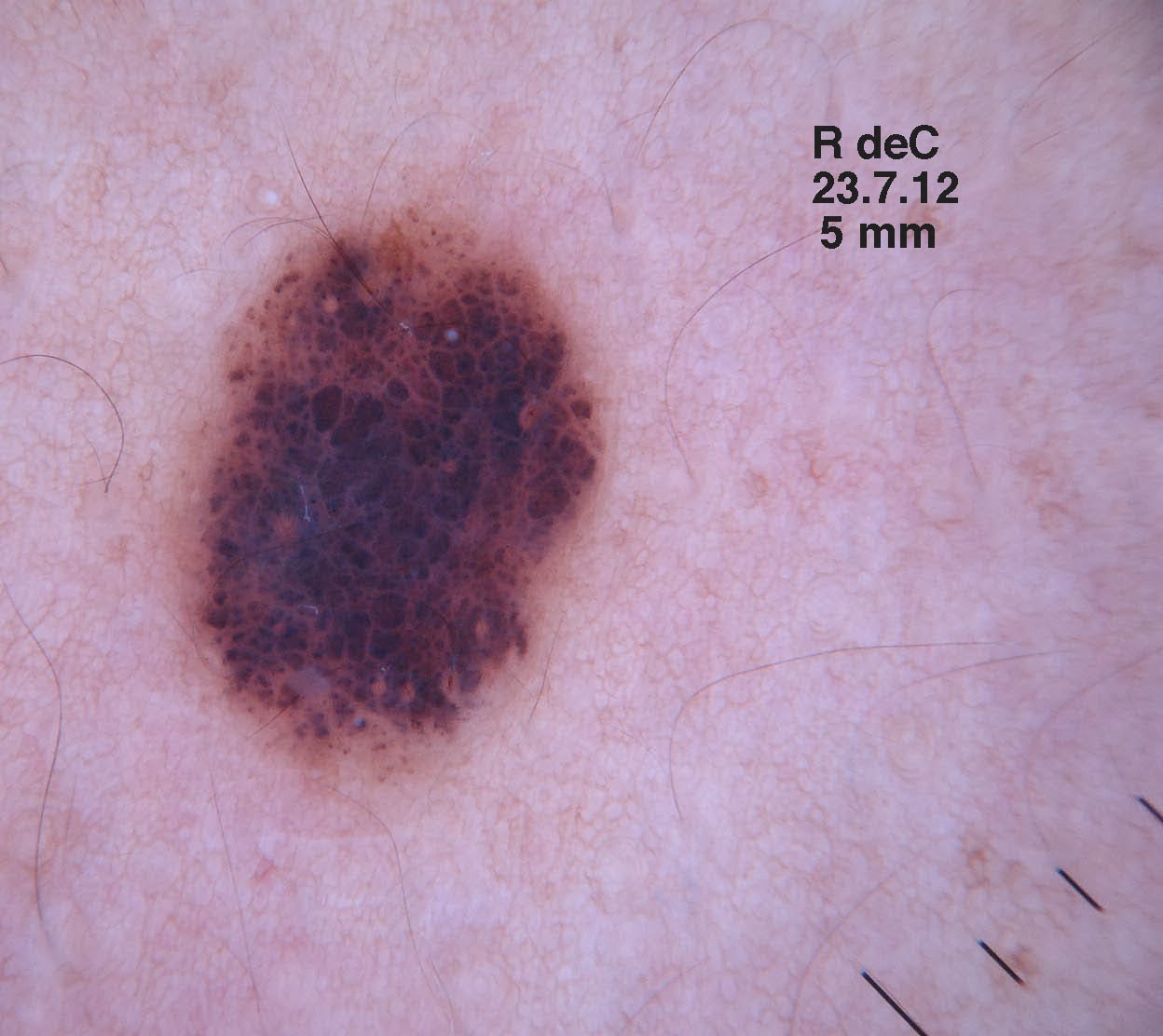
Clinical Photos:


Lab:  CBC, chemistries normal.  IgE 867 IU/Ml

Pathology:  Initial bx signed out as SCC.  Since he has scores of lesions repeat biopsies of an early and more developed lesion were taken.

Diagnosis:

Discussion and Questions:

References:
1. Journal of the American Academy of Dermatology
Volume 69, Issue 3 , Pages 426-430, September 2013
Keratoacanthomas arising in association with prurigo nodules in pruritic, actinically damaged skin
Timothy P. Wu, BA, Kristen Miller, MD, David E. Cohen, MD, Jennifer A. Stein, MD, PhD  jennifer.Stein@nyumc.org

2. J Clin Pharm Ther. 2013 Feb;38(1):16-8. doi: 10.1111/jcpt.12005. Epub 2012 Sep 26.
Treatment of prurigo nodularis with pregabalin.
Mazza M, Guerriero G, Marano G, Janiri L, Bria P, Mazza S.

3. Dermatol Ther. 2010 Mar-Apr;23(2):194-8. doi: 10.1111/j.1529-8019.2010.01314.x.
Therapeutic hotline: Treatment of prurigo nodularis and lichen simplex chronicus with gabapentin.
Gencoglan G, Inanir I, Gunduz K.  (no real data on patient background)

4. Eur J Dermatol. 2008 Jan-Feb;18(1):85-6. Epub 2007 Dec 18.
Gabapentin for the treatment of recalcitrant chronic prurigo nodularis.
Dereli T, Karaca N, Inanir I, Oztürk G.  Available Free Full Text.

Saturday, February 1, 2014

Changing Nevus

Presented by:
Dr. Salvatore Donatello
Dermatologica e Venereologia. Catania, Sicilia


Abstract:  44 year old man with changing naevus

HPI:  The patient was seen in July of 2012 for a general cutaneous exam. He has Type IV skin. A naevus was noted on his right shoulder.  Dermatoscopic exam was felt to be not particularly worrisome but he was asked to return to have the lesion rechecked in six months.  He returned 18 months later.  The patient thought it had been present for many years and had not noted change.

O/E: July 2012:  5 mm in diameter dark brown papule  on right shoulder.  At the time, I thought the dermatoscopic exam looked normal.  It did look like an active lesions

January 2014:  The lesion is still 5 mm in diameter.  However, the brown clods noted on dermatoscopy in 2012 have disappeared and the lesion now looks uniformly dark gray.

Clinical photos:

Clinical Photo 31.1.14
Dermatoscopic Photo 31.1.14
Plan:  The lesion was excised with 3 mm margins on 31.1.14

Follow-up:  The biopsy report showed that this is an intradermal melanocytic without any atypical features. The brown clods in the first biopsy most likely are a sign of an active growing lesion.

Discussion:  Neither lesion on its own was very worrisome.  However, fortunately we have dermatoscopic images from July 2012 and January 2014.  The brown clods have disappeared and the lesion looks uniformly gray. Although the patient was asked to return in six months he did not do so.  We have no way of knowing if the change would have been noted then.
The pathology will tell us the story.  Is this an evolving naevus or a melanoma.  The practice of medicine can be humbling. There are few articles on evolving naevi in the literature.  This case will teach us something.

We will add an update after the biopsy is signed out. I have sent the specimen to a dermatologist in Napoli with a special interest in pigmented lesions.