This is default featured slide 1 title

Go to Blogger edit html and find these sentences.Now replace these sentences with your own descriptions.

This is default featured slide 2 title

Go to Blogger edit html and find these sentences.Now replace these sentences with your own descriptions.

This is default featured slide 3 title

Go to Blogger edit html and find these sentences.Now replace these sentences with your own descriptions.

This is default featured slide 4 title

Go to Blogger edit html and find these sentences.Now replace these sentences with your own descriptions.

This is default featured slide 5 title

Go to Blogger edit html and find these sentences.Now replace these sentences with your own descriptions.

Showing posts with label Dermoscopy. Show all posts

Wednesday, February 27, 2013

Dermatoscopy of Molluscum

The patient, a two year old girl, was referred for evaluation of a 5 mm in diameter tumor on the left shoulder present for a few months.  The lesion was a dome-shaped papule that was in the presence of numerous smaller but similar lesions (the latter were typical molluscum).

Dermoscopy of molluscum has been described in the literature.  The most salient feature is presence of polylobular amorphous white to yellowish globules as seen in the larger of the two lesions below.  The smaller lesion shows just a solitary amorphous lobule.


Discussion:  Dermatoscopy is a useful tool for the diagnosis of molluscum.  The presence of the white globules (which represent the molluscum bodies) is a pathognomonic sign.  In this large lesion, multiple molluscum bodies are the tip-off to the diagnosis.

Cliff Rosendahl writes: “This is a non-pigmented lesion, circular in shape with a sharply demarcated border over the total periphery. Centrally there are white clods and white structureless areas on a pink background with serpentine vessels.  In ‘Dermatoscopy’ page 236 the characterisation is “White to yellow clods or structureless zone and curved vessels at the periphery which do not cross the centre” so this example is a variation of that description.”


References:
1. An Bras Dermatol. 2011 Jan-Feb;86(1):74-9.
Dermoscopic patterns of molluscum contagiosum: a study of 211 lesions confirmed by histopathology.  Free full text
Abstract
RESULTS: At clinical examination and dermoscopy of 211 lesions, orifices were visualized in 50.24% and 96.68% of the lesions, and vessels in 6.16% and 89.10%, respectively. The vascular patterns found in the 188 lesions in which vessels were found at dermoscopy were the crown (72.34%), radial (54.25%) and punctiform patterns (20.21%). Half of the 188 lesions had a combination of vascular patterns, with the flower pattern (a new vascular pattern) being found in 19.68% of cases. More orifices and vessels were identified at dermoscopy than at clinical examination, including cases with inflammation or perilesional eczema and small lesions. Punctiform vessels were associated with inflammation, excoriation and perilesional eczema.
CONCLUSIONS: Dermoscopy performed on molluscum contagiosum lesions proved superior to dermatological examination even in cases in which clinical diagnosis was difficult. The presence of orifices, vessels and specific vascular patterns aids diagnosis, including differential diagnosis with other types of skin lesion.  

2.  Arch Dermatol. 2005 Dec;141(12):1644.  Dermoscopy of molluscum contagiosum.
Morales A, Puig S, Malvehy J, Zaballos P.

Saturday, December 15, 2012

Lichen Nitidus

Presented by Yoon Cohen, D.O.

Abstract: 13 year-old boy with a few years history of lichen nitidus

HPI:  This is a 13 year-old Hispanic boy who presented for treatment of warts. During the exam, multiple small white shiny dots were noticed. The patient has been aware of these asymptomatic lesions for the past few years.  He had been given triamcinolone 0.1% ointment bid, but forgot to apply it because it did not bother him. 

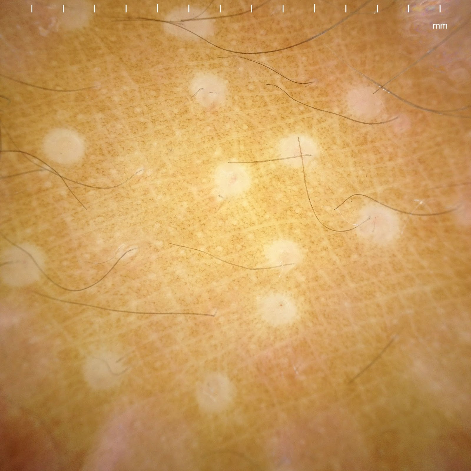
O/E:  There  are multiple pinhead-sized 1 mm white shiny flat-topped papules in an oval shape on the right knee.  

Dermatoscopic Photo:


Multiple small white circles
The dermoscopic image taken with the Canfield Dermatoscope with iPhone attachment

Diagnosis:

Lichen nitidus

Discussion:
While there are many dermoscopic studies on the skin cancers, there is a growing interest of dermoscopic features on other skin diseases such as inflammatory diseases, connective tissue disorders, or even psychogenic related skin diseases. Lichen nitidus is easily recognized clinically with experienced eyes. With my own interest in dermoscopy, I report an interesting dermoscopic finding of lichen nitidus. There are 1-2 mm multiple small white circles which represent subepidermal infiltrate of lymphocytes, histiocytes, and multinucleated giant cells in the dermal papillae. 

Lichen nitidus was first described by Pinkus in 1907. Lichen nitidus, which means shiny papules, is a relatively uncommon, asymptomatic, chronic eruption, consisting of minute sharply demarcated skin-colored papules. It has a predilection for males (4:1) and the mean age of onset was 7 yrs for males and 13 yrs for females. Typically, patients present for assessment with an asymptomatic or mildly pruritic eruption. The eruption may be localized to one or more areas or generalized in distribution. The pathogenesis of lichen nitidus is unknown. It was originally believed to be a form of tuberculid and more recently considered to be a variant of lichen planus. Histologially, lichen nitidus is characterized by a circumscribed collection of inflammatory cells in the papillary dermis that abuts the overlying epidermis. The inflammatory infiltrate consists of lymphocytes, histiocytes, and multinucleated giant cells. DIF negative.  Most cases require no treatment due to the asymptomatic nature of the eruption and tendency for spontaneous resolution. For symptomatic cases, moderately potent or potent topical steroid therapy may help. There are a number of anecdotal reports of improvement or clearance with narrow band UVB phototherapy, extensive sunlight exposure, oral astemizole, oral cetirizine-levamisol combination, topical dinitrochlorobenzene, itraconazole and oral cyclosporin. Oral retinoids have been used successfully in the treatment of palmoplantar lichen nitidus.

Reference:
Schachner LA, Hansen RC. Pediatric Dermatology. 4th edition. 2011
L. Nitidus eMedicine (free open access)

Comment: By DJ Elpern.  This is the first report of the dermatoscopic appearance of L. nitidus.  Yoon Cohen deserves credit for this observation.


Wednesday, April 25, 2012

Dermoscopy Rocks

Presented by Hamish Dunwoodie, MBBS
Locum Tenens Physician,
Dakota First Nation, Portage La Prairie, Manitoba

Overview:  The patient is an otherwise healthy artist from Moncton, New Brunswick who is studying native pottery production with a First Nation's band in Manitoba.  She presented to our clinic with a one week history of a black macule on the left thenar eminence.  The patient has a past history of nonmelanoma skin cancer and is worried that this may be a melanoma.

O/E:  There is a 1 mm black macule in the above-mentioned area.  Dermoscopic image suggests a vascular lesion.  Note reddish black globules.

The lesions was shaved off, a small amount of H2O2 was applied and the residual hemorrhagic area was digested. The lesion disappeared!

Photographs:  Clinical Image, Dermatoscopic Image before shaving. Dermatoscopic image after shave and H2O2.


Discussion:  Dermoscopy established that this is a vascular lesion, a "subcorneal haematoma."  Once the dermatoscopic image was appreciated, it was gently shaved off with a # 15 scalpel and it vanished.  End of story.  We needed no special tools in this isolated practice setting to put the patient's mind at ease.  I am sure that some of our dermatoscopy experts will have more to day.

Reference:
Zalaudak I, et. al.  Dermoscopy of subcorneal hematoma. Dermatol Surg  Dermatol Surg. 2004 Sep;30( 9):1229-32.
Abstract
BACKGROUND:
Subcorneal hematoma is a pigmented skin lesion usually occurring on palms or soles after a trauma or sport activity. Clinically, it may exhibit overlapping features with acral melanoma or acral melanocytic nevi, leading to unnecessary excision of this otherwise harmless skin lesion.
OBJECTIVE:
The objective was to describe the dermoscopic features in a series of subcorneal hematomas.
METHODS:
Dermoscopic images of 15 subcorneal hematomas were evaluated for the presence of different colors and dermoscopic structures.
RESULTS:
In our series, a red-black hue was the most frequent color seen by dermoscopy (40% of the lesions) and a homogeneous pattern of pigmentation was the most frequent dermoscopic structure (53.3%). Remarkably, 40% of the lesions exhibited a parallel-ridge pattern that is usually found in early melanoma of palms and soles. In 46.7% of the lesions, red-black globules were additionally seen at the periphery as satellites disconnected from the lesion's body. Only two lesions showed either parallel-furrow or fibrillar pattern. A scratch test performed in four lesions, allowed complete or partial removal of the pigmentation.
CONCLUSION:
Dermoscopic features of subcorneal hematomas may be similar to those observed in acral melanocytic lesions. Nevertheless, in most cases the correct diagnosis can be facilitated by the presence of a red-black homogeneous pigmentation, often combined with satellite globules. A positive scratch test may be considered as an additional diagnostic clue.


Sunday, October 17, 2010

The Power of BLINCK

Presented by Yoon Cohen MS IV, University of New England, Biddeford, Maine and David Elpern MD, Williamstown, Massachusetts

Abstract: 68 yo woman with 4-6 months history of an atypical melanocytic lesion.

HPI: This healthy 68 yo woman with type II skin presented to the clinic with 4-6 months history of an atypical melanocytic lesion on the left knee. She had noticed an increase in size and change in color and was concerned about these changes in the lesion.

O/E: There was a 7 mm in a diameter asymmetrical brownish macule on the left knee. The lesion showed an irregular border with varied colors.

Clinical photographs:


Dermoscopic images:


Microscopic images:
Dermatopathology report:
The specimen exhibits a proliferation of moderate to severely atypical melanocytes distribubted in irregular nests, as well as singly at and above the dermal epidermal junction, with pagetoid spread to the granular layer and near confluence over at least three rete ridges. These findings support the histologic diagnosis of melanoma-in-situ.

Our appreciation to Dr. Deon Wolpowitz, MD from Boston Univeresity, Dermatopathology, for providing these photomicrographs for the case.

4X


10X


20X


20X


Diagnosis:
Malignant melanoma in situ

Discussion:

The BLINCK approach:

We followed the BLINCK checklist, introduced by Dr. Peter Bourne, a founder of the Skin Cancer College of Australia and New Zealand (SCCANZ). The score for the lesion was added up to 4 by criteria as following.

1. B. The lesion was not clearly benign at our first initial evaluation
2. L. The lesion appeared to be lonely without any other similar melanocytic lesion near by
3. I. The lesion appeared to be irregular outline and color on our dermoscopic exam
4. N & C. The patient was nervous about the change in color in past 4-6 months
5. K. The lesion exhibited known clues when viewed with a dermatoscope. See "Chaos and Clues" reference below.
BLINCK Score = 4

According to the BLINCK approach, a lesion should be biopsied if the BLINCK score is 2 or more out of a possible 4. Therefore, the we excised this lesion and sent for a pathologic evaluation.

According to Dr. Bourne, the BLINCK approach is presented as a simple method to assist the clinician with the decision of whether to biopsy a skin lesion or not. The use of this algorithm will improve the pickup rate of potentially serious skin cancers as well as reduce the number of unnecessary benign lesion excisions. BLINCK may be especially helpful to clinicians who have only basic or intermediate dermoscopy skills but who are regularly called upon to assess skin lesions in their practices.

Questions:
1. Would you consider using the BLINCK approach at your practice?
2. If you already have adapted the BLINCK approach, how have your experiences been?

References:
1. McColl I. BLINCK. http://idsblinck.blogspot.com/2009/11/blinck.html. Updated November 19, 2009. Accessed September 7, 2010
2. Rosendahl C, Kittler H, Cameron A, et al. CHAOS & CLUES - The Algorithm. http://www.chaosandclues.blogspot.com. Updated November 19, 2009. Accessed September 7, 2010