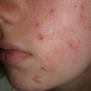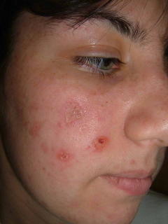

A repeat biopsy on June 27. 2005 showed:
DIAGNOSIS: Skin - Left Temple:
Epidermal necrosis with s cale crust containing neutrophils , sub-epidermal abundant neutrophils and fibrin deposition, superficial and deep perivascular lymphohistiocytic infiltrate with focal neutrophil ic microabscesses, septal and lobular panniculitis with mixed inflammatory cell infiltrate of abundant neutrophils , lymphocytes , histiocytes, and eosinophils and numerous activated endothelial cells, surrounding a medium-sized vessel with marked mixed inflammatory cell infiltrate of neutrophils , histiocytes and occasional eosinophils .
NOTE : These changes are suggestive of a medium-sized vasculitis with overlying necrosis. Elastic tissue stain (EVG) does not reveal the vessel in the deeper sections, therefore, arterial or venular distinction cannot be made. The differential diagnosis includes a large vessel vasculitis such as periarteritis nodosa or early Wegener's granulomatosis. P.A.S. stain is negative for fungal organisms. Fite stain is negative for mycobacteria . However, an infectious vasculitis cannot be entirely excluded . If the clinical suspicion persists, culture studies may be of help . The differential diagnosis also includes , in the appropriate clinical setting , factitial panniculitis with secondary vascular involvement. These are not the changes of lupus erythematosus , pityriasis lichenoides et varioliformis acuta or prurigo nodularis . Serologic studies may be helpful. Clinico-pathologic correlation is suggested.


0 comments:
Post a Comment